This whole rabbit serum was prepared by repeated immunizations with a synthetic peptide corresponding to amino acid residues of human Notch 1 located near the N-terminal sequence of the cleaved N intracellular domain (NICD).
Konjugation:
Unconjugated
Alternative Synonym:
rabbit anti-notch1 antibody, Neurogenic locus Notch homolog protein 1, hN1, Translocation-associated Notch protein TAN-1
0.02 M Potassium Phosphate, 0.15 M Sodium Chloride, pH 7.2
Formulierung:
Liquid (sterile filtered)
Target-Kategorie:
Human
Antibody Type:
Primary Antibody
Application Verdünnung:
ELISA: 1:20,000 - 1:60,000, IHC: 1:200, IF Microscopy: User Optimized, IP: User Optimized, WB: 1:500- 1:2,000
Anwendungsbeschreibung:
Anti-Notch 1 has been tested by ELISA, dot blot, western blot and immunohistochemistry. An 80 kDa band corresponding to Notch 1 was observed at a 1:500 dilution. Specific conditions for reactivity should be optimized by the end user.
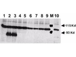
Rabbit anti-Human NOTCH 1 (Cleaved N Terminal) was used at a 1:500 dilution to detect mouse Notch 1 by Western blot. Equivalent amounts of lysates from transiently transfected 293 cells expressing recombinant myc-tagged mouse Notch constructs were electrophoresed and transferred to membrane using standard methods. A reaction with diluted primary antibody was followed by washing, reaction with a 1:10,000 dilution of HRP conjugated Gt-a-Rabbit IgG (611-103-122), and color development. Lane M: Mol wt markers. Lane 1: No transfection. Lane 2: N1 (mouse deleted extracellular domain)-myc. Lane 3: N1 (mouse intracellular domain)-myc. Lane 4: N2 (mouse deleted extracellular domain)-myc. Lane 5: N2 (mouse intracellular domain)-myc. Lane 6: N3 (mouse deleted extracellular domain)-myc. Lane 7: N3 (mouse intracellular domain)-myc. Lane 8: N4 (mouse deleted extracellular domain)-myc. Lane 9: N4 (mouse intracellular domain)-myc. Lane 10: N1 (mouse deleted extracellular domain)(V to G)-myc. Personal communication, Dr. Stacey Huppert.
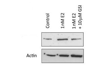
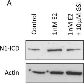
Western Blot of Rabbit anti-Notch1 antibody. Lane 1: MCF-7 control lysate. Lane 2: MCF-7 +1 nM 17beta-estradiol. Lane 3: MCF-7 + 10 µM gamma secretase inhibitor. Load: 35 µg per lane. Primary antibody: Notch1 antibody at 1:500 for overnight at 4C. Secondary antibody: IRDye800(TM) rabbit secondary antibody at 1:10,000 for 45 min at RT. Block: 5% BLOTTO overnight at 4C. Predicted/Observed size: 80 kDa for Notch1.
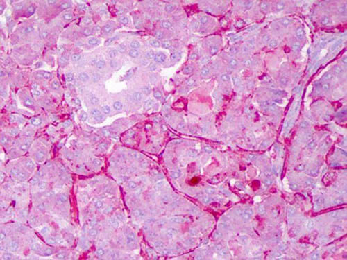
Immunohistochemistry of Rabbit anti-Notch1 antibody. Tissue: Exocrine glands of human pancreas. Fixation: FFPE. Primary antibody: Notch1 antibody at 1:200. Staining: moderate to strong membranous staining and faint to moderate cytoplasmic staining. Islets showed faint staining.
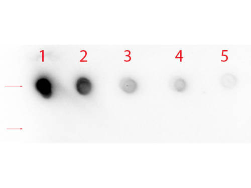
Dot Blot of Rabbit anti-Notch 1 (Cleaved N Terminal) (Human Specific) Antibody. Antigen: Row 1 - Notch 1 Peptide (Cleaved N Terminal) Row 2 - Notch 1 (Intra) Peptide. Load: Lane 1 - 200 ng Lane 2 - 66.67 ng Lane 3 - 22.22 ng Lane 4 - 7.41 ng Lane 5 - 2.47 ng. Primary antibody: Rabbit anti-Notch 1 (Cleaved N Terminal) (Human Specific) Antibody at 1:1,000 for 60 min at RT. Secondary antibody: HRP Rabbit Secondary at 1:40,000 for 30 min at RT. Block: MB-070 for 1 HR at RT.
Systemic oestrogen signalling is mediated by EGFR and Notch. (A) Representative Western blot showing expression of cleaved (active) Notch1 (N1-ICD) following culture 1 nM 17beta-estradiol 10 µM GSI. (Bi) Representative Western blot showing expression of Notch ligands in sorted MCF7 cells (left) and, where available, metastatic cells (right). (Bii) Densitometric analysis of three independent repeats of MCF7 sorting and of a single experiment for primary cells. Comparisons between population 1 (CSC enriched) and other populations are displayed. (C and D) Mammosphere formation was assessed following culture with 1 nM 17beta-estradiol gamma secretase inhibitor (GSI) alone and in combination with gefitinib. Fold change is normalised to control, untreated cells represented as line. (E) Representative image of protein levels of ERK and phosphorylated (actived) ERK following culture for 48 hours in monolayer 10 µM GSI. Means plotted SEM, *P < 0.05, **P < 0.01, ***P < 0.001 compared to E2 treated. P < 0.05 compared to control cells. Figure provided by CiteAb. Source: Breast Cancer Res, PMID: 23497505.
* Mehrwertsteuer und Versandkosten nicht enthalten. Irrtümer und Preisänderungen vorbehalten