This antibody was prepared from whole rabbit serum produced by repeated immunizations with a synthetic peptide corresponding to a region near the C-terminal of mouse PI3K p110d. This sequence is identical in both mouse and human.
0.02 M Potassium Phosphate, 0.15 M Sodium Chloride, pH 7.2
Formulierung:
Liquid (sterile filtered)
Target-Kategorie:
Mouse
Antibody Type:
Primary Antibody
Application Verdünnung:
ELISA: 1:4,000 - 1:20,000, WB: 1:5,000
Anwendungsbeschreibung:
Anti-PI3 kinase p110 delta subunit antibody has been tested for use in ELISA, western blotting and immunoprecipitation. Reactivity in other immunoassays is unknown. A mouse whole cell splenic lysate is suitable for use as a positive control.
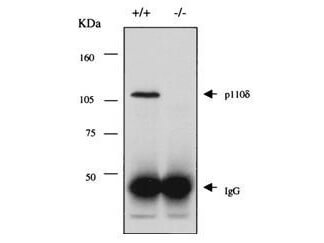
Immunoprecipitation and western blot using ROCKLAND Immunochemicals Rabbit-anti-PI3K p110d antibody. Lane 1 shows the detection of a single band corresponding to mouse p110d detected in a lysate from p110d +/+ mice (lane 1) and the absence of staining in a similar lysate isolated from p110d -/- mice (lane 2). Molecular weight markers confirm a MW of ~120 kDa. In both instances 2 mg of a total splenic lysate was used for immunoprecipitation and western blot analysis. For IP use ~5 µl of antiserum. For WB use a 1:5,000 dilution of antiserum. Detection occurs using a 1:2,000 dilution of HRP Goat-a-Rabbit IgG (611-103-122) with visualization via ECL. Film exposure approximately 45. Other detection systems will yield similar results. See Jou et al for additional details.
* Mehrwertsteuer und Versandkosten nicht enthalten. Irrtümer und Preisänderungen vorbehalten