The immunogen is a Red Fluorescent Protein (RFP) fusion protein corresponding to the full length amino acid sequence (234aa) derived from the mushroom polyp coral Discosoma.
Konjugation:
Unconjugated
Alternative Synonym:
goat anti-RFP antibody, DsRed, rDsRed, Discosoma sp. Red Fluorescent Protein, Red fluorescent protein drFP583
0.02 M Potassium Phosphate, 0.15 M Sodium Chloride, pH 7.2
Formulierung:
Liquid (sterile filtered)
Antibody Type:
Primary Antibody
Application Verdünnung:
ELISA: 1:2,000 - 1:10,000, IF Microscopy: User Optimized, WB: 1:1000-1:10,000
Anwendungsbeschreibung:
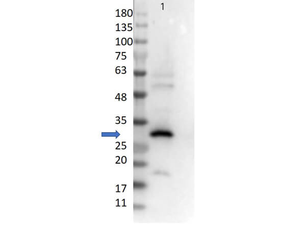
Polyclonal anti-RFP has been tested by western blot and is designed to detect RFP and its variants. Optimal titers for applications should be determined by the researcher. Expect a band ~27kDa in Western Blot in the appropriate cell lysate or extract.
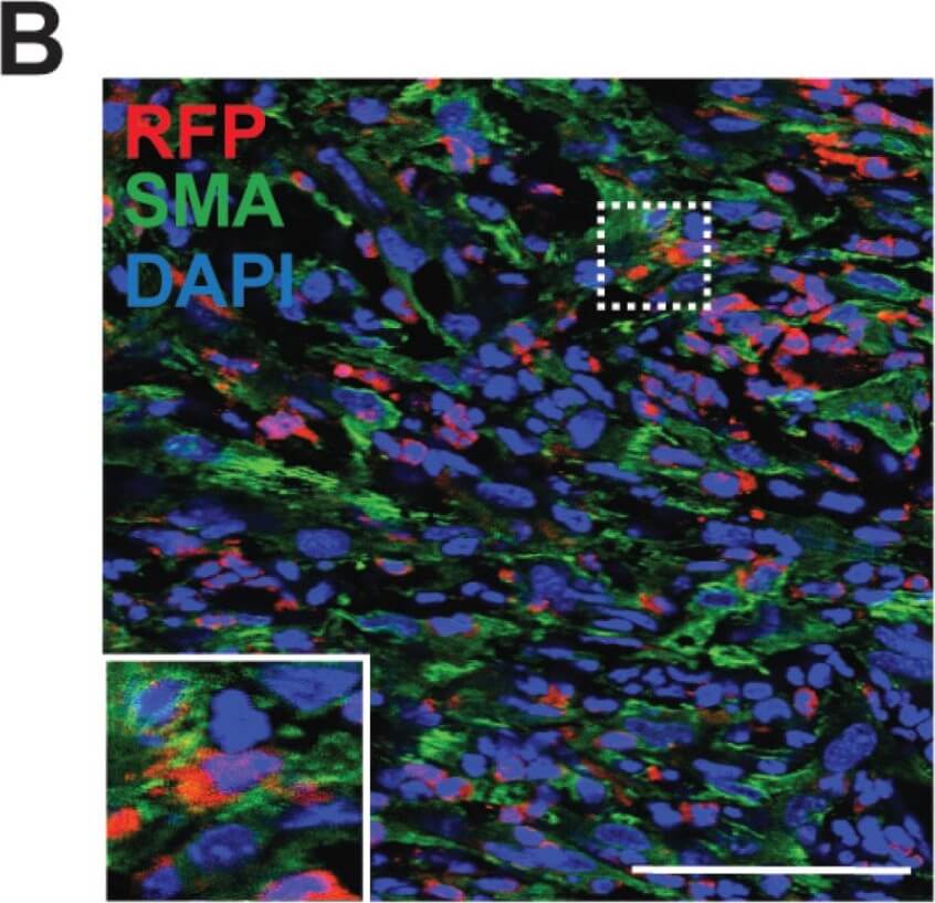
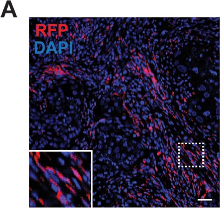
Population of CD11b + myeloid progenitor cells differentiate into SMA + stromal cells within tumors and in vitro. (A) Representative image of red fluorescent protein (RFP) + stromal cells in tumor from CCR2-RFP heterozygous SCID mouse. (B) RFP + SMA + double positive cells within tumor stroma. (C) CD11b + SMA + double positive cells within tumor stroma. (D) CD45 + CD11b + CD34+ myeloid progenitor cells in the mammary gland at 1.5 (n = 5 empty vector (EV), 7 CCL2) and 2.5 weeks (n = 5 EV, 7 CCL2) post-transplantation were quantified by flow cytometry. (E) CD45 + CD11b + CD34 + myeloid progenitor cells in the bone marrow at 1.5 weeks (n = 6 EV, 7 CCL2) and 2.5 weeks (n = 3 mice/group) post-transplantation were quantified by flow cytometry. (F) Representative brightfield image of colony formed by CD45 + CD11b + CD34 + myeloid progenitor cells isolated using fluorescence-activated cell sorting (FACS). (G) Colonies in culture co-stained with SMA and collagen I. Statistical differences determined by Mann-Whitney U test. Magnification bars = 50 µm. Figure provided by CiteAb. Source: Cancers (Basel), PMID: 32731354.
Western Blot of Goat Anti-RFP Antibody. Lane 1: 50ng of RFP protein (p/n 000-001-379). Primary Antibody: Goat Anti-RFP at 1:10,000 overnight at 2-8C. Secondary Antibody: Donkey anti-Goat IgG HRP (p/n 605-703-125) at 1:40,000 for 30 min at RT. Blocking buffer: BlockOut (p/n MB-073). Predicted MW: ~27kDa.
Population of CD11b + myeloid progenitor cells differentiate into SMA + stromal cells within tumors and in vitro. (A) Representative image of red fluorescent protein (RFP) + stromal cells in tumor from CCR2-RFP heterozygous SCID mouse. (B) RFP + SMA + double positive cells within tumor stroma. (C) CD11b + SMA + double positive cells within tumor stroma. (D) CD45 + CD11b + CD34+ myeloid progenitor cells in the mammary gland at 1.5 (n = 5 empty vector (EV), 7 CCL2) and 2.5 weeks (n = 5 EV, 7 CCL2) post-transplantation were quantified by flow cytometry. (E) CD45 + CD11b + CD34 + myeloid progenitor cells in the bone marrow at 1.5 weeks (n = 6 EV, 7 CCL2) and 2.5 weeks (n = 3 mice/group) post-transplantation were quantified by flow cytometry. (F) Representative brightfield image of colony formed by CD45 + CD11b + CD34 + myeloid progenitor cells isolated using fluorescence-activated cell sorting (FACS). (G) Colonies in culture co-stained with SMA and collagen I. Statistical differences determined by Mann-Whitney U test. Magnification bars = 50 µm. Figure provided by CiteAb. Source: Cancers (Basel), PMID: 32731354.
* Mehrwertsteuer und Versandkosten nicht enthalten. Irrtümer und Preisänderungen vorbehalten