0.02 M Potassium Phosphate, 0.15 M Sodium Chloride, pH 7.2
Formulierung:
Liquid (sterile filtered)
Antibody Type:
Primary Antibody
Application Verdünnung:
ELISA: 1:200 - 1:1000, IHC: 1:1500, IF Microscopy: User Optimized, WB: 1:1000 - 1:5000
Anwendungsbeschreibung:
Anti-Luciferase Antibody has been tested in Western Blot, IHC, IF, and ELISA. Expect a band ~60kDa in appropriate cell lysates. Although not tested, this antibody would be useful in immunoprecipitation, immunocytochemistry, and most immunological methods
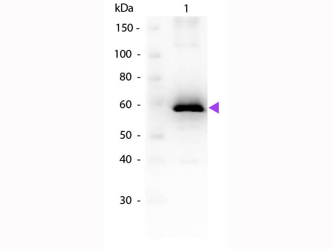
Western Blot of Goat anti-Luciferase Antibody. Lane 1: Luciferase. Load: 50ng per lane. Primary antibody: Luciferase antibody at 1:1000 overnight at 4C. Secondary antibody: Peroxidase goat secondary antibody (p/n 611-103-122) at 1:40,000 for 30 min at RT. Block: (p/n MB-070) for 30 min at RT. Predicted/Observed size: ~60 kDa for Luciferase. Other band(s): None.
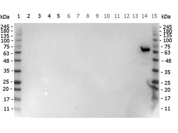
Western Blot of Goat anti-Luciferase antibody. Lane 1 Marker: Opal Pre-stained ladder (p/n MB-210-0500). Lane 2: HEK293 lysate (p/n W09-000-365). Lane 3: HeLa Lysate (p/n W09-000-364). Lane 4: CHO/K1 Lysate (p/n W07-000-357). Lane 5: E-coli HCP Control (p/n 000-001-J08). Lane 6: J774A.1 Lysate (p/n W10-001-GX3). Lane 7: C2C12 Lysate (p/n W10-001-GL7). Lane 8: Mouse Embryonic Fibroblast Lysate (p/n W10-001-371). Lane 9: NIH/3T3 Lysate (p/n W10-000-358). Lane 10: Mouse Liver Lysate (p/n W10-000-T020). Lane 11: PC-12 Lysate (p/n W12-001-GL9). Lane 12: Rat Brain Lysate (p/n W12-000-T077). Lane 13: Rat Testis Lysate (p/n W12-000-GZ3). Lane 14: Luciferase [Photinus pyralis (Firefly)]. Lane 15 Marker: Opal Pre-stained ladder (p/n MB-210-0500). Load: 10 µg of lysate or 50ng of purified protein per lane. Primary antibody: Luciferase antibody at 1:1,000 overnight at 4C. Secondary antibody: Peroxidase Goat secondary antibody (p/n 605-703-125) at 1:40,000 for 60 min at RT. Blocking Buffer: 1% Casein-TTBS (MB-082) for 30 min at RT. Predicted/Observed size: ~60 kDa for Luciferase.
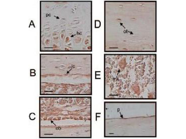
Luciferase immunostaining was analyzed in tibias from ovx ERE-luciferase mice taken 24 h after a 17-E2 injection. Positive luciferase staining was identified in hypertrophic chondrocytes (hc) (A), lining cells (lc) (B), osteoblasts (ob) (C), a subpopulation (10%) of osteocytes (ot) (upper arrow points at a positively stained osteocyte, whereas the lower depicts a negative osteocyte) (D), and megakaryocytes (E). Faint staining was found on the periosteal surface (p) (F). No background staining was seen when omitting the primary antibody (data not shown). The bar in the lower left corner represents 25 µm. pc, Proliferative chondrocyte.
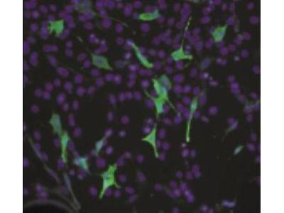
NIH3T3 cells transiently transfected with a luciferase gene. Luciferase-positive cells were detected using Rocklands polyclonal Anti-Luciferase antibody. Cells (25,000/well) were transiently transfected with a pLuc plasmid. After 2 days, the cells were fixed using 4% paraformaldehyde, permeabilized with 0.1% Triton X-100, and blocked with 1% normal donkey serum. Cells were stained with 20µg/ml Anti-Luciferase in PBS for 2 hours followed by 1:200 dilution of donkey anti-goat IgG-FITC (green) for 1 hour. Cells were mounted using Vectashield with DAPI (blue) and visualized at 200X magnification with a fluorescent microscope.
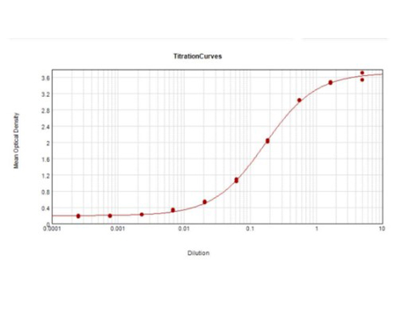
ELISA Results of Goat Anti-Luciferase Antibody. Each well was coated in duplicate with 1.0 µg of Luciferase [Firefly]. The starting dilution of antibody was 5µg/ml and the X-axis represents the Log10 of a 3-fold dilution. The titer is 1:6,050. This titration is a 4-parameter curve fit where the IC50 is defined as the titer of the antibody. Assay performed using TMB substrate p/n TMBE-1000.
* Mehrwertsteuer und Versandkosten nicht enthalten. Irrtümer und Preisänderungen vorbehalten