![]()
|
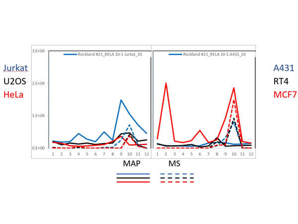
PAGE-MAP (microsphere affinity proteomics) of Mouse Anti-NFKB p65 (Rel A) Antibody. (Catalog Number: 200-301-065, Lot Number: 26076). Antibody array western blot binding of gelfree size separated fractions of multiple lysates (solid lines) and shotgun mass spectroscopy identification (dashed lines) of the target band run in parallel correlate confirming the specificity of this antibody against NFKB p65. Data was provided by the Lund-Johansen lab of Oslo University Hospital. For more information on PAGE-MAP/IP-MS identification of antibody specificity and its large-scale implementation for |
![]()
|
|
![]()
|
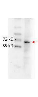
Rockland anti NFKB p65 (Rel A) monoclonal antibody (p/n 200-301-065) was used to detect ~65 kD band (red arrow) in HeLa whole cell lysate (p/n W09-000-364). Lysate was run on 4-20% gradient gel transferred under standard conditions and blocked in 1% BSA-TBST for 30 min at RT. Blot was probed with monoclonal anti-NFkB p65 at 1:1000 in 1% BSA-TBST o/n at 4C and detected with HRP conjugated Rb-anti-Mouse antibody (p/n 610-4302) at 1:40,000 in (p/n MB-070) for 30 min at RT. |
![]()
|
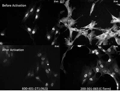
Anti NFkB monoclonal antibody - Immunocytochemistry. Tissue: Human Fibroblasts. Top: Before activation. Bottom: After activation with poly IC. Left: Anti-p65 NLS specific - (p/n 600-401-271, lot 18372). Right: Anti-p65 C-Term monoclonal antibody - (p/n 200-301-065, lot 26076). The two antibodies that are shown target different regions of the p65 protein. The different staining patterns are thought to correspond with different functional regions of the protein. |
![]()
|
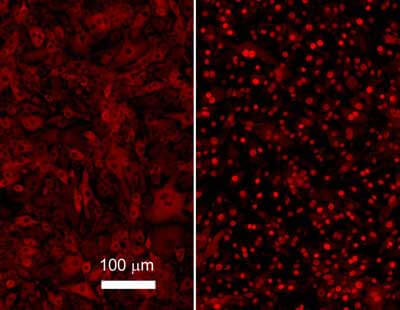
Rockland Monoclonal anti NFKB p65 (Rel A) antibody was used to detect p65 in b.end5 mouse endothelial cells. Unstimulated control cells (left) show cytoplasmic staining, TNF-alpha stimulated cells (right) show nuclear staining. For staining, cells were washed with PBS to remove all traces of culture media and fixed with paraformaldehyde 4 % (45 min). Slides were washed with PBT (phosphate buffer 0.1M + Triton-X-100 0.1 %), 3 times 10 min each, then, blocked with PBT + 5 % normal goat serum, 1 hour. Sample was Incubated overnight in primary antibody (1:600 in blocking buffer). After 3X wash in PBT for 10 min, slides were incubated 1 hour with secondary antibody (1:1000) and mounted in 1:1 PB glycerol. Images kindly provided by Tebu-Bio from Francisco Javier Carrillo-Salinas (PhD Student), Dra. Carmen Guaza Instituto Cajal (CSIC), Madrid, Spain. |
![]()
|
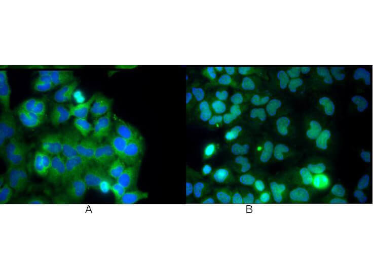
Rockland Monoclonal anti NFKB p65 (Rel A) antibody was used to detect p65 by immunofluorescence at a dilution of 1:5000. Hela cells were grown to sub-confluent on 18 mm2 glass coverslips 1.5. Cells were either unstimulated (A), or stimulated (B) with 50 ng/ml of TNF alpha for 30 min prior fixation. Cells were then fixed in methanol and blocked with 10% normal goat serum (NGS), in PBS, and TritonX 0.2% (Tx) and incubated for 1 hr at RT with primary ab, counterstained with DAPI and washed in PBS/NGS/Tx. Cells were incubated for 1 hr at RT with Atto 425 conjugated anti mouse secondary antibody for STED CW imaging. Data was collected on a STED-CW TCS-SP5 Confocal system equipped with a DFC 350FX camera allowing sequential acquisition in widefiled, confocal and STED CW imaging on the same system. |
![]()
|
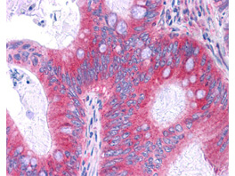
Rockland Antibody (p/n 200-301-065) has been tested in immunohistochemistry, analyzed by an anatomic pathologist and validated for use in IHC applications against formalin-fixed, paraffin-embedded human tissues. Showed moderate to strong staining within many tissues, including epithelium of the breast, small intestine, kidney, pancreas, prostate, skin, placenta, and uterus, as well as within neurons and lymphoid tissues such as spleen, thymus, and tonsil. The antibody produced an excellent signal with almost no background staining at a concentration of 2.5µg/ml. The image displayed shows specific staining in colon carcinoma as the precipitated red signal, with a hematoxylin purple nuclear counterstain. Image provided courtesy of LifeSpan Biosciences, Seattle, WA. |