This antibody was produced in mesothelin-deficient mice by immunizations with plasmid cDNA encoding human MSLN full length protein followed by a single boost of a recombinant human mesothelin-Fc fusion protein.
This antibody has been tested for use in immunohistochemistry, flow cytometry, and western blotting. Specific conditions for reactivity should be optimized by the end user. Expect a band approximately 40 kDa in size corresponding to mature mesothelin by
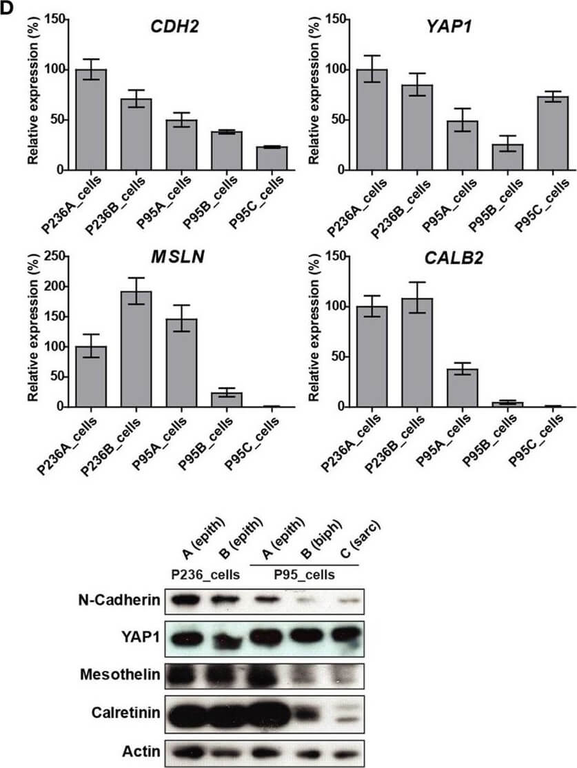
(A) Tumor processing flow-chart of 235 samples, which were processed for RNA extraction, cell culture and embedding either in PFA or OCT. Samples showing attributes which are grayed in the chart, were not used in the live cell biobank. (B) Circos whole genome copy number variations (CNVs) view of tumor and primary cell culture in patients malignant pleural mesothelioma (MPM) 236 and MPM95. The quilt plot highlights the CNV and SNVs in genes that are part of MPM landscape (2). (C) Immunofluorescence analysis of selected markers in primary culture from patient MPM236. Scale bar 200µm. (D) Selected genes expression analysis at mRNA (upper panel) and protein (lower panel) level in primary cultures derived from samples from two patients, MPM236 and MPM95. The latter one underwent EMT during disease progression. (E) Selected genes expression analysis at mRNA in tumor samples from patients MPM236 and MPM95. (F) Significant correlation between gene expression changes in tumor and primary culture from patient MPM236 at passage 3. Figure provided by CiteAb. Source: Front Oncol, PMID: 29527515.
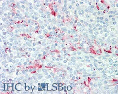
Immunohistochemistry of Mouse anti-Mesothelin antibody. Tissue: human tonsil. Fixation: formalin fixed paraffin embedded. Antigen retrieval: not required. Primary antibody: anti-Mesothelin antibody at 15 µg/mL for 1 h at RT. Secondary antibody: Peroxidase mouse secondary antibody at 1:10,000 for 45 min at RT. Staining: Mesothelin as precipitated red signal with hematoxylin purple nuclear counterstain.
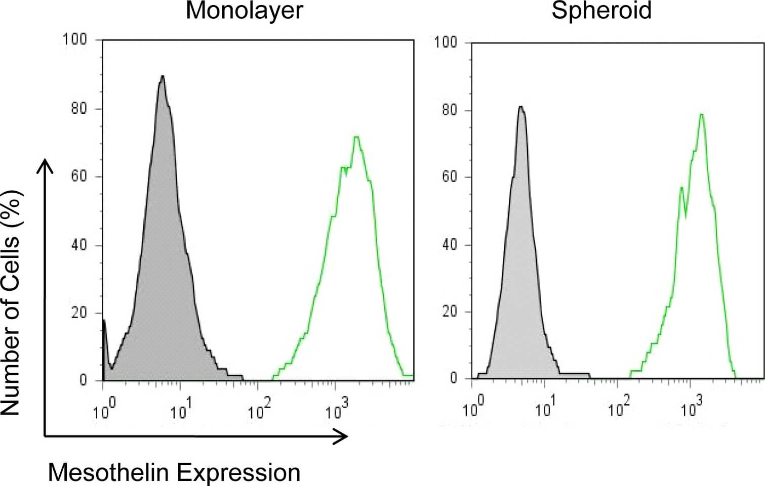
Mesothelin expression in mesothelioma monolayers and spheroids.NCI-H226 cells incubated with an anti-mesothelin mAb (MN) and detected with goat anti-mouse IgG conjugated with Alexa488 by flow cytometry. Figure provided by CiteAb. Source: PLoS One, PMID: 21305058.
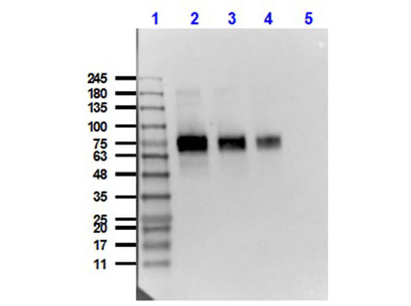
Western Blot of Mouse Anti-Mesothelin Antibody. Lane 1: Opal Prestained Molecular Weight Marker (p/n MB-210-0500). Lane 2: HeLa [10µg] + Mesothelin-Fc [0.1µg]. Lane 3: HeLa [10µg] + Mesothelin-Fc [0.05µg]. Lane 4: HeLa [10µg] + Mesothelin-Fc [0.02µg]. Lane 5: HeLa Whole Cell Lysate (p/n W09-000-364). Primary Antibody: Anti-Mesothelin at 1µg/mL overnight at 2-8C. Secondary Antibody: Rabbit Anti-Mouse IgG HRP conjugated (p/n 610-4302) at 1:40,000 for 30 mins at RT. Block: BlockOut Buffer (p/n MB-073) 30 mins at RT. Exposure: 15 sec. Predicted MW: 40kDa Mesothelin + Fc region 30kDa. Observed MW: ~70-75kDa.
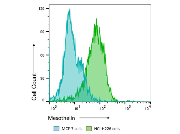
Flow Cytometry Results of Anti-Mesothelin (MOUSE) Monoclonal Antibody. The green histogram shows NCI-H226 cells and blue histogram shows MCF-7 cells. Both cell lines are stained with a 1:800 dilution Anti-Mesothelin (MOUSE) Monoclonal Antibody. The secondary antibody use was Anti-Mouse IgG (H&L) (GOAT) Antibody DyLight(TM) 488 Conjugated (p/n 610-141-002, lot43322) at the 1:400 dilution.
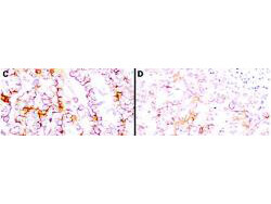
Immunohistochemistry using Rocklands anti-mesothelin antibody to react with two epitopes on mesothelin in PEFF human mesothelioma tissue sections treated by antigen retrieval methods. Anti-mesothelin primary antibodies were used at 10 µg/mL to label these sections as follows: C, MAb MB, and D, MAb MN followed by goat anti-mouse IgG conjugated to horseradish peroxidase at 25 µg/mL in 1% BSA/PBS for 30 minutes. (magnification, *200, bar, 50 µm). Reprinted with permission from Clin.Cancer Res. 11(16):5840-6.
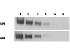
Western blotting using Rocklands anti-mesothelin antibodies to detect mesothelin-Fc at 100 ng (lane 1), 25 ng (lane 2), 6 ng (lane 3), 2 ng (lane 4) and 0.4 ng (lane 5). Lane 6 contains 50 ng of CDC25-Fc. Proteins were separated on 4-20% gradient gel by SDS-PAGE followed by transfer to PVDF membrane. Primary antibody was used at 1 µg/ml followed by reaction with ALP goat anti-mouse IgG and BCIP/NBT substrate. Reprinted with permission from Clin.Cancer Res. 11(16):5840-6.
* Mehrwertsteuer und Versandkosten nicht enthalten. Irrtümer und Preisänderungen vorbehalten