This Protein G purified monoclonal antibody was prepared using conventional hybridoma technology after repeated immunizations with 3-(4-Hydroxy-3-nitrophenyl acetamido) propionic acid - BSA conjugate.
This Protein G purified antibody has been tested for use in ELISA, immunoprecipitation, immunohistochemistry and western blotting. Specific conditions for reactivity should be optimized by the end user. Approximately 0.7 µg of antibody is sufficient to d
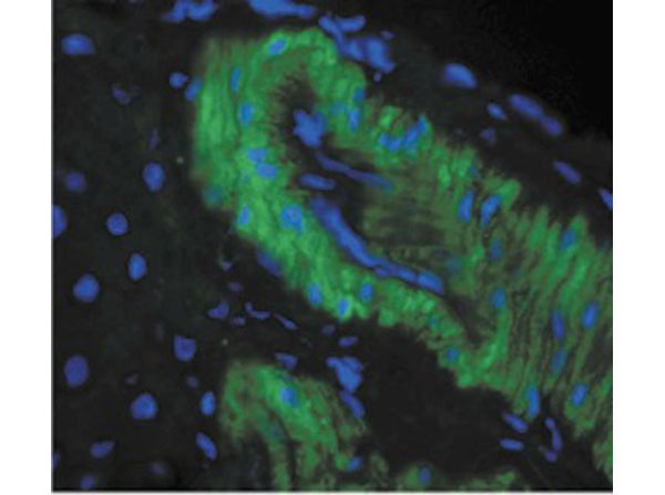
Immunohistochemistry of Mouse Anti-Nitrotyrosine Monoclonal Antibody. Tissue: rat liver tissue. Primary Antibody: Mouse Anti-Nitrotyrosine Monoclonal Antibody at 1:1000. Secondary Antibody: FITC Goat Anti-Mouse (green).
Immunohistochemistry of Mouse Anti-Nitrotyrosine Monoclonal Antibody. Tissue: mouse inflamed colon. Fixation: Formalin with anti-microbial. Primary Antibody: Mouse Anti-Nitrotyrosine Monoclonal Antibody at 1:1,000,000 for 12 hours at 4C. Secondary Antibody: Biotin Goat Anti-Mouse at 1:2000 for 1 hour at RT. Counterstain: Mayer Hematoxylin (purple/blue) nuclear stain at 200 µl for 2 minutes at RT. Magnification: 40x. This image was produced using an amplifying IHC wash buffer. The antibody has therefore been diluted more than is recommended for other applications.
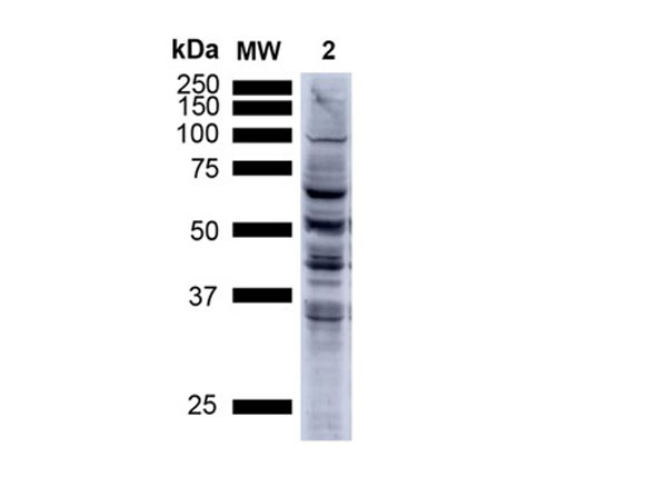
Western Blot of Anti-Nitrotyrosine. Load: 15 ug. Lane 1: MW ladder. Lane 2: Human A549 Cells. Block: 5% Skim Milk in TBST. Primary Antibody: Mouse Anti-Nitrotyrosine Monoclonal Antibody at 1:1000 for 2.5 hours at RT. Secondary Antibody: Goat anti-mouse IgG HRP at 1:1000 for 1 hour at RT. Color Development: Chemiluminescent for HRP for 5 min in RT. Predicted/Observed Size: Multiple Bands.
Immunohistochemistry of Mouse Anti-Nitrotyrosine Monoclonal Antibody. Tissue: mouse backskin from transgenic mice. Fixation: Bouins Fixative and paraffin-embedded. Primary Antibody: Mouse Anti-Nitrotyrosine Monoclonal Antibody at 1:100 for 1 hour at RT. Secondary Antibody: FITC Goat Anti-Mouse (green) at 1:50 for 1 hour at RT.
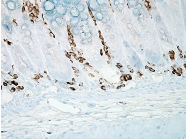
Immunohistochemistry of Mouse Anti-Nitrotyrosine Monoclonal Antibody. Tissue: human colon carcinoma. Fixation: Formalin. Primary Antibody: Mouse Anti-Nitrotyrosine Monoclonal Antibody at 1:25,000 for 12 hours at 4C. Secondary Antibody: Biotin Goat Anti-Mouse at 1:2000 for 1 hour at RT. Counterstain: Mayer Hematoxylin (purple/blue) nuclear stain at 200 µl for 2 minutes at RT. Magnification: 40x.
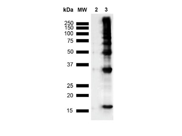
Western Blot of Anti-Nitrotyrosine. Lane 1: MW Ladder. Lane 2: hASYN Monomer (3.84 ug). Lane 3: Nitrosylated hASYN (3.84 ug). Block: 5% Skim Milk in TBST. Primary Antibody: Mouse Anti-Nitrotyrosine Monoclonal Antibody at 1:1000 for 2 hours at RT. Secondary Antibody: Goat anti-mouse IgG HRP at 1:4000 for 2 hour at RT. Color Development: Chemiluminescent for HRP for 5 min in RT. Predicted/Observed Size: Multiple Bands.
Western Blot of Anti-Nitrotyrosine. Lane 1: MW Ladder. Lane A: Nitrosylated-HEK293 (15uL). Lane B: HEK293 (15 ug). Block: 5% Skim Milk in TBST. Primary Antibody: Mouse Anti-Nitrotyrosine Monoclonal Antibody diluted in 1.5% BSA and TBST for 1 hours at RT. Secondary Antibody: Goat anti-mouse IgG HRP at 1:4000 for 1 hour at RT. Predicted/Observed Size: Multiple Bands.
* Mehrwertsteuer und Versandkosten nicht enthalten. Irrtümer und Preisänderungen vorbehalten