Anti-Rhodopsin antibody has been tested in Western Blot and Immunohistochemistry. Expect a band of approximately 39 kDa in size corresponding to the rhodopsin proteins in Western blot in the appropriate cell lysate or extract. Researchers should determin
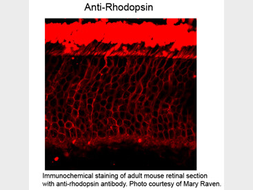
Immunohistochemistry of Mouse anti-Rhodopsin antibody. Tissue: mouse retinal. Fixation: formalin fixed paraffin embedded. Antigen retrieval: not required. Primary antibody: Rhodopsin antibody at 1:100 for 1 h at RT. Secondary antibody: Peroxidase mouse secondary antibody at 1:10,000 for 45 min at RT. Localization: Rhodopsin is in the rod spherules. Staining: Rhodopsin as precipitated red signal.
* Mehrwertsteuer und Versandkosten nicht enthalten. Irrtümer und Preisänderungen vorbehalten