Cav1.3 Antibody was produced in mice by repeated immunizations with a fusion protein corresponding to an internal region of rat Cav1.3.
Konjugation:
Unconjugated
Alternative Synonym:
CACH3, CACN4, CACNA 1D, CACNL1A2, voltage dependent L type calcium channel subunit alpha 1D, Calcium channel, L type, alpha-1 polypeptide, isoform 2, Rat brain class D
0.02 M Potassium Phosphate, 0.15 M Sodium Chloride, pH 7.2
Formulierung:
Liquid (sterile filtered)
Target-Kategorie:
Rat
Antibody Type:
Primary Antibody
Application Verdünnung:
IHC: 0.1-1.0ug/mL, IF Microscopy: 1.0-10ug/mL, IP: User Optimized, WB: 1-10ug/mL
Anwendungsbeschreibung:
Anti-Cav1.3 Antibody was tested in WB, IP, IHC, and IF microscopy. Expect a band approximately ~250kDa on specific lysates. Specific conditions for reactivity should be optimized by the end user.
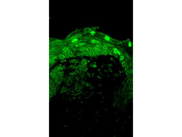
Immunofluorescence Microscopy of mouse anti-CAV 1.2 antibody. Tissue: Human hippocampus tissues. Fixation: N/A. Primary antibody: anti-CAV 1.2 for 1h at RT. Secondary antibody: Fluorescein mouse secondary antibody at 1:10,000 for 45 min at RT. Localization: Membrane. Staining: Cav 1.2 as precipitated green signal.
* Mehrwertsteuer und Versandkosten nicht enthalten. Irrtümer und Preisänderungen vorbehalten