Anti-Calbindin-D is tested for Immunofluorescence, Immunohistochemistry -P and Western Blot. Specific conditions for reactivity should be optimized by the end user. Expect a band approximately ~30 kDa corresponding to the appropriate cell lysate or extra
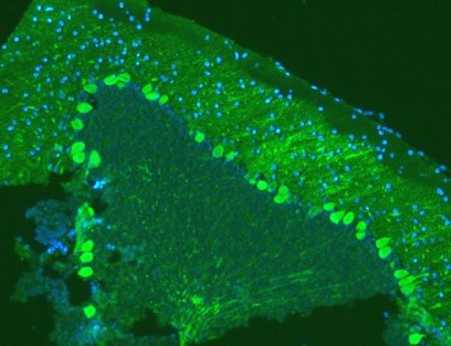
Immunofluorescence analysis of Calbindin-D using anti-Calbindin-D antibody. Calbindin-D was detected in paraffin-embedded section of mouse brain tissues. Heat mediated antigen retrieval was performed in citrate buffer (pH6, epitope retrieval solution) for 20 mins. The tissue section was blocked with 10% goat serum. The tissue section was then incubated with 1µg/mL mouse anti-Calbindin-D Antibody overnight at 4C. DyLight488 Conjugated Goat Anti-Mouse IgG secondary antibody at 1:100 dilution for 30 minutes at 37C. The section was counterstained with DAPI. Visualize using a fluorescence microscope and filter sets appropriate for the label used.
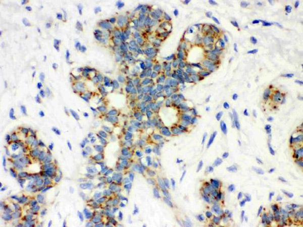
Immunohistochemistry of Mouse Anti-Calbindin-D antibody.Tissue: Human Mammary Cancer Tissue. IHC(P).
Western blot analysis of Calbindin-D using anti-Calbindin-D antibody. Electrophoresis was performed on a 5-20% SDS-PAGE gel at 70V (Stacking gel) / 90V (Resolving gel) for 2-3 hours. The sample well of each lane was loaded with 50µg of sample under reducing conditions. Lane 1: rat brain tissue lysates, Lane 2: rat kidney tissue lysates, Lane 3: mouse brain tissue lysates. After Electrophoresis, proteins were transferred to a nitrocellulose membrane at 150mA for 50-90 minutes. Blocked the membrane with 5% Non-fat Milk/TBS for 1.5 hour at RT. The membrane was incubated with affinity purified monoclonal mouse anti-Calbindin-D antigen primary antibody at 0.5 µg/mL overnight at 4C, then washed with TBS-0.1% Tween 3 times with 5 minutes each and probed with a goat anti-mouse IgG-HRP secondary antibody at 1:10,000 for 1.5 hour at RT. The signal is developed using an Enhanced Chemiluminescent detection (ECL) kit with Tanon 5200 system. A specific band was detected for Calbindin-D at approximately 28KD. The expected band size for Calbindin-D is at 28KD.
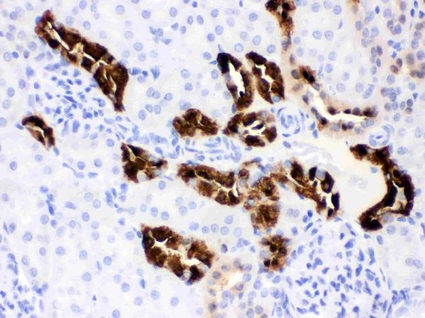
Immunohistochemistry of Mouse Anti-Calbindin-D antibody.Tissue: Mouse Kidney Tissue. IHC (P).
Immunohistochemistry of Mouse Anti-Calbindin-D antibody.Tissue: Human Intestinal Cancer Tissue. IHC (P).
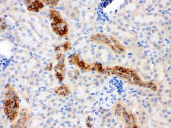
Immunohistochemistry of Mouse Anti-Calbindin-D antibody.Tissue: Rat Kidney Tissue. IHC (P).
* Mehrwertsteuer und Versandkosten nicht enthalten. Irrtümer und Preisänderungen vorbehalten