Anti-Calponin has been tested in Immunohistochemistry -P, Immunocytochemistry, and Western Blot. Specific conditions for reactivity should be optimized by the end user. Expect a band approximately ~33 kDa corresponding to the appropriate cell lysate or e
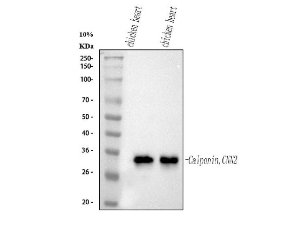
Western blot analysis of Calponin/CNN2 using anti-Calponin/CNN2 antibody. Electrophoresis was performed on a 5-20% SDS-PAGE gel at 70V (Stacking gel) / 90V (Resolving gel) for 2-3 hours. The sample well of each lane was loaded with 30 µg of sample under reducing conditions. Lane 1: chicken heart tissue lysates, Lane 2: chicken heart tissue lysates. After electrophoresis, proteins were transferred to a nitrocellulose membrane at 150 mA for 50-90 minutes. Blocked the membrane with 5% non-fat milk/TBS for 1.5 hour at RT. The membrane was incubated with affinity purified mouse anti-Calponin/CNN2 antigen monoclonal antibody at 0.5 µg/mL overnight at 4C, then washed with TBS-0.1%Tween 3 times with 5 minutes each and probed with a goat anti-mouse IgG-HRP secondary antibody at 1:10,000 for 1.5 hour at RT. The signal is developed using an Enhanced Chemiluminescent detection (ECL) kit with Tanon 5200 system. A specific band was detected for Calponin/CNN2 at approximately 33 kDa. The expected band size for Calponin/CNN2 is at 33 kDa.
* Mehrwertsteuer und Versandkosten nicht enthalten. Irrtümer und Preisänderungen vorbehalten