Anti-5-mC Antibody was produced in mice by repeated immunization with 5-methylcytosine.
Konjugation:
Unconjugated
Alternative Synonym:
5-methylcytosine, 5-methyl cytosine
Klonalität:
Monoclonal
Konzentration:
1.24 mg/ml by UV absorbance at 280 nm
Klon-Bezeichnung:
[33D3]
Isotyp:
IgG1
Puffer:
0.01 M Sodium Phosphate, 0.25 M Sodium Chloride, pH 7.2
Formulierung:
Liquid (sterile filtered)
Target-Kategorie:
Human
Antibody Type:
Primary Antibody
Application Verdünnung:
ELISA: 1:3,000, ChIP: 1 µg/ChIP, WB: 1:1,000
Anwendungsbeschreibung:
Anti-5-mC Antibody is tested for Dot Blotting, ELISA, Immunofluorescence, Methylated DNA Immunoprecipitation (MeDIP), and Sequencing (MeDIP-seq). Specific conditions for reactivity should be optimized by the end user.
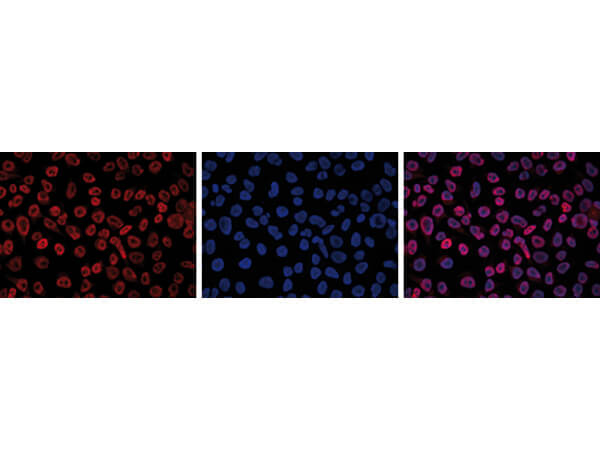
Immunofluorescence Microscopy results of Mouse anti-5-mC antibody. Tissue: HeLa cells. Fixation: 4% formaldehyde in PBS for 10 min at RT. Retrieval: 0.5% Triton X-100 for 1 hr, treated with 2N HCl for 1 hr. Block: PBS/0.1% Triton X-100/1% BSA. Primary antibody: 5-mC antibody at 1:500 for 1 hr at RT (left). Secondary antibody: Goat anti-Mouse Alexa594 secondary antibody at 1:10,000 for 45 min at RT. Staining: 5-mC antibody as red fluorescent signal (left), DAPI blue (middle), merge of the two stainings (right).
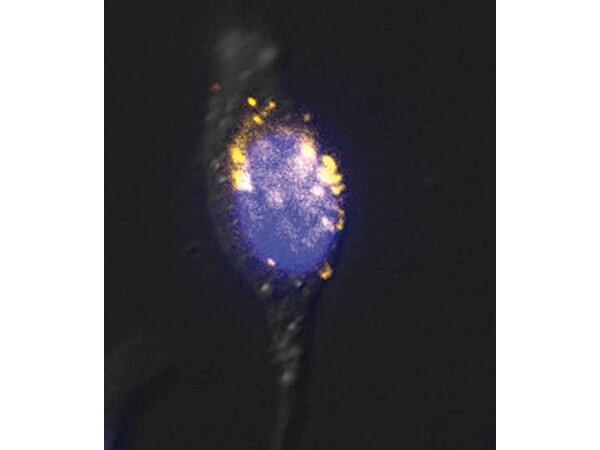
Immunofluorescence Microscopy results of Mouse anti-5-mC antibody. Tissue: Interphase HeLa cell. Fixation: 4% formaldehyde in PBS for 10 min at RT. Retrieval: 0.5% Triton X-100 for 1 hour and treated with 2N HCl for 1 hour. Block: PBS/0.1% TritonX-100/1% BSA. Primary antibody: 5-mC antibody at 1:500 for 1 hr at RT. Secondary antibody: Goat anti-Mouse FITC secondary antibody at 1:10,000 for 45 min at RT. Staining: : 5-mC antibody as yellow fluorescent signal with Hoescht staining (blue).
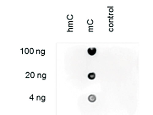
Dot Blot results of Mouse anti-5-mC monoclonal antibody. Antigens: hmC, mC, and, C control forms of the immunizing peptide. Load: 100 ng, 20 ng, and 4 ng as indicated. Primary antibody: 5-mC antibody at 1:250 for 45 min at 4C. Secondary antibody: Dylight(TM)488 anti-mouse secondary antibody at 1:10,000 for 45 min at RT. Block: 5% BLOTTO overnight at 4C.
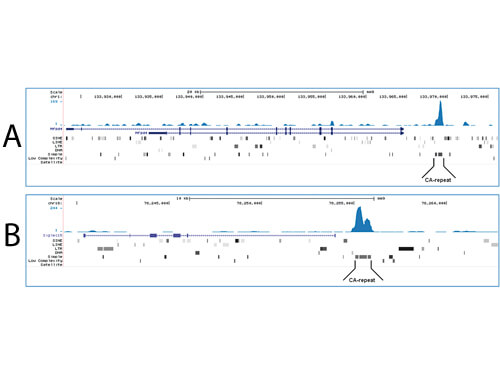
Methylated DNA immunoprecipitation sequencing with anti-5-mC Antibody. Genomic DNA from E14 ES cells were sheared to generate random fragments (size range 300 to 700 bp). One µg of the fragmented DNA was ligated to Illumina adapters and the resulting DNA was used for a standard MeDIP assay, using 2 µg of the 5-mC. Figure A and B show Genome browser views of CA simple repeat elements with read distributions specific for 5-mC at 2 gene locations (SigleC15 and Mfsd4). Visual inspection of the peak profiles in a genome browser reveals high enrichment of CA simple repeats in affinity-enriched methylated fragments after MeDIP with the Mouse Anti-5-mC Antibody.
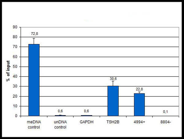
Methylated DNA Immunoprecipitation (MeDIP) was performed on fragmented human genomic DNA using the monoclonal antibody against 5-mC. The fragmented DNA was spiked with controls methylated DNA (meDNA) as a positive and unmethylated DNA (unDNA) as a negative control) prior to performing the IP. QPCR was performed with primer sets and for a known methylated (TSH2B) and unmethylated (GAPDH) genomic region. An additional internal positive and negative control locus (4994+ and 8804-, respectively) were also tested (4994+: forward primer 5-GGGAATATAAGGAGCGCACA-3 and reverse primer 5- TCGGTTAAAACGGTCAGGTC-3, 8804-: forward primer 5-CGAGGCGTGAGTTATTCCTG-3 and reverse primer 5-CTCTTGTGGCTGAGCTCCTT-3). This figure shows the recovery (mean of 3 experiments), expressed as a % of input (the relative amount of immunoprecipitated DNA compared to input DNA after qPCR analysis).
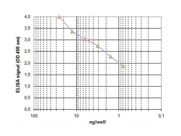
ELISA results of mouse anti-5-mC antibody. ELISA was performed using monoclonal antibody against 5-mC diluted 1:100. The wells were coated with a serial dilution of the methylated DNA control.
* Mehrwertsteuer und Versandkosten nicht enthalten. Irrtümer und Preisänderungen vorbehalten