Anti-AKT1 Antibody was produced in mice by repeated immunizations with a synthetic peptide corresponding to internal residues of human AKT1 protein followed by monoclonal development.
0.02 M Potassium Phosphate, 0.5 M Sodium Chloride, pH 7.2
Formulierung:
Lyophilized
Target-Kategorie:
Human
Antibody Type:
Primary Antibody
Application Verdünnung:
ELISA: User Optimized, Flow Cytometry: User Optimized, IHC: User Optimized, IF Microscopy: User Optimized, WB: User Optimized
Anwendungsbeschreibung:
Anti-AKT1 FITC Antibody has been tested by ELISA and western blot and is suitable for Flow Cytometry, immunohistochemistry, and western blotting. Expect a band approximately 56 kDa in size corresponding to AKT1 protein by western blotting in the appropri
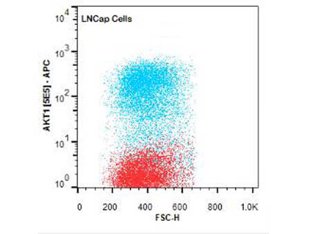
Flow Cytometry of Mouse anti-AKT1 antibody. Cells: LNCap Cells. Stimulation: none. Primary antibody: Allophycocyanin AKT1 antibody at 1.0 µg/mL for 20 min at 4C.
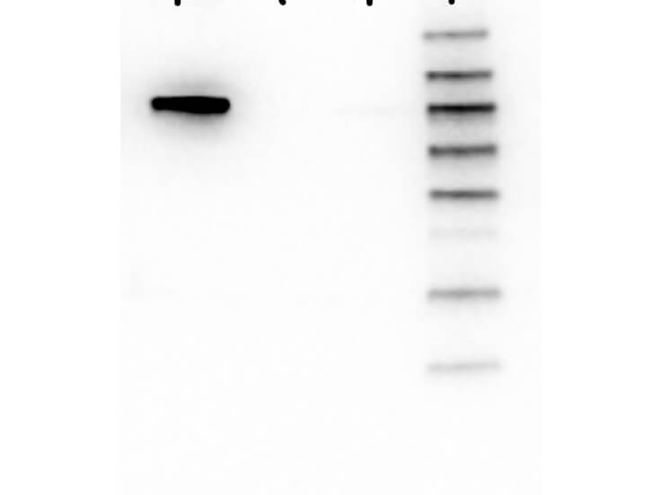
Western Blot of Mouse anti-AKT1 antibody. Lane 1: GST-AKT1. Lane 2: GST-AKT2. Lane 3: GST-AKT3. Lane 4: Molecular Weight Marker. Load: 25 ng per lane. Primary antibody: AKT1 antibody at 1:1000 for overnight at 4C. Secondary antibody: Mouse secondary antibody at 1:40,000 for 30 min at RT. Block: 5% BLOTTO overnight at 4C. Predicted/Observed size: 78 kDa for AKT1. Other band(s): none.
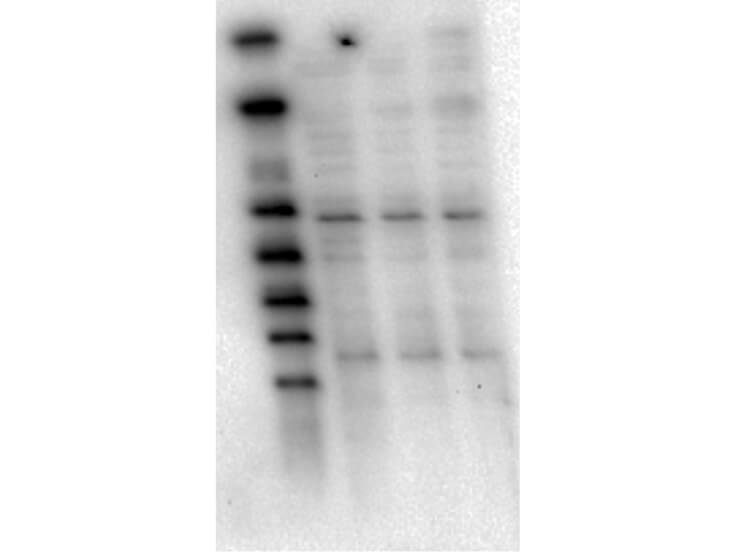
Western Blot of Mouse Anti-AKT1 antibody. Lane 1: Molecular Weight Marker. Lane 2: LnCap lysate (p/n W09-001-GJ9). Lane 3: Jurkat lysate (p/n W09-001-370). Lane 4: MDA-MB 468 lysate (p/n W09-001-GG9). Load: 5 µg per lane. Primary antibody: AKT1 antibody at 1:1000 for overnight at 4C. Secondary antibody: Mouse secondary antibody at 1:20,000 for 45 min at RT. Block: 5% BLOTTO overnight at 4C. Predicted/Observed size: 56 kDa for AKT1.
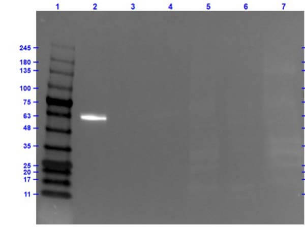
Western Blot of Mouse Anti-AKT1 Fluorescein Conjugated Antibody.Lane 1: Opal Prestained Molecular Weight Marker (p/n MB-210-0500). Lane 2: AKT1 Recombinant Protein (0.05µg). Lane 3: AKT2 Recombinant Protein (0.05µg). Lane 4: AKT3 Recombinant Protein (0.05µg). Lane 5: HEPG2 Whole Cell Lysate (20µg). Lane 6: C2C12 Whole Cell Lysate (20µg). Lane 7: A549 Whole Cell Lysate (20µg). Primary Antibody: Mouse Anti-AKT1 FITC at 1.0µg/mL overnight at 2-8C. Secondary Antibody: Rabbit Anti-Mouse HRP (p/n 610-403-C46) at 1:40,000 for 30mins at RT.Blocking: Fluorescent Buffer (p/n MB-073) for 1hr at RT.Predicted MW: ~56kDa. Exposure: 0.5 sec.
* Mehrwertsteuer und Versandkosten nicht enthalten. Irrtümer und Preisänderungen vorbehalten