This protein A purified Jagged 1 antibody was prepared from whole rabbit serum produced by repeated immunizations with a synthetic peptide corresponding to amino acids near the N-terminus of human Jagged-1 protein.
Anti-Jagged-1 antibody has been tested for use in ELISA, immunohistochemistry, immunofluorescence microscopy and western blot. Specific conditions for reactivity should be optimized by the end user. Expect a band approximately 150 kDa in size correspondi
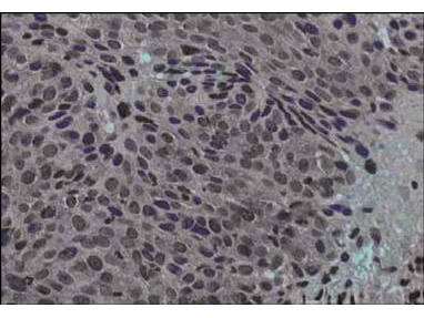
Immunohistochemical staining of human cervical cancer tissue (40X magnification) using Rocklands Protein A purified anti-Jagged-1 antibody. Tissue was fixed with formalin and embedded in paraffin. Hematoxylin was used to counter-stain cells. A 1:100 dilution of primary antibody was used. Personal Communication. Martin Kast Laboratory.
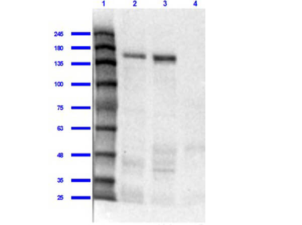
Western Blot of Rabbit Anti-Jagged 1 Antibody. Lane 1: Opal Prestained MW marker (p/n MB-210-0500). Lane 2: Mouse Liver Whole Cell Lysate [10µg] (p/n W10-000-T020). Lane 3: Human Liver Whole Cell Lysate [10µg]. Lane 4: Human Lung Whole Cell Lysate [10µg]. Primary Antibody: Anti-Jagged 1 at 1:1000 overnight at 2-8C. Secondary Antibody: Goat Anti-Rabbit IgG Peroxidase Conjugated (p/n 611-103-122) at 1:70000 for 30mins at RT. Blocking Buffer: BlockOut Buffer (p/n MB-073) for 1hr RT. Predicted Molecular Weight: 113kDa. Exposure: 30 sec.
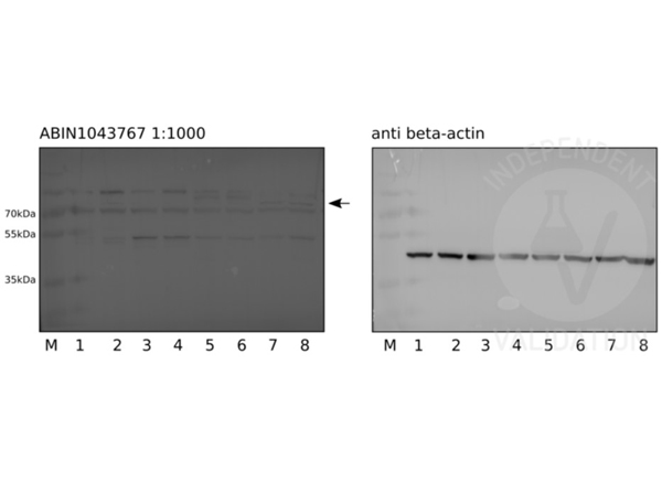
Western blot Anti-Jagged-1 (RABBIT) Antibody. Human pluripotent stem cells (lanes 1 and 2), definite endoderm cells (lanes 3 and 4), pancreatic endoderm cells (lanes 5 and 6), and pancreatic progenitors (lanes 7 and 8). Primary Antibody: Anti-Jagged-1 used at a 1:1000 (30sec exposure) overnight at 4C. Beta-actin served as loading control at 1:2000 at RT for 1hr. Secondary Antibody: donkey anti-rabbit HRP conjugated antibody or donkey anti-mouse HRP conjugated antibody diluted 1:5000 for 1h at RT. Independently Validated byantibodies-online GmbH (p/n ABIN1043767/ ABIN129524) courtesy of Ulm University Hospital.
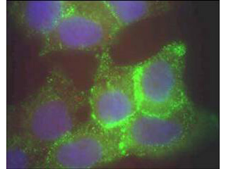
Immunofluorescence microscopy using Rocklands Protein A purified anti-Jagged-1 antibody of human corneal epithelial cells. Primary antibody was used at a 1:500 dilution. The Jagged1 (green staining) is localized to the cytoplasm and is consistent with reports in the literature. The nucleus is stained with Bis benzamine (blue). Personal Communication. Aihua Ma, Univdersity of Cardiff.
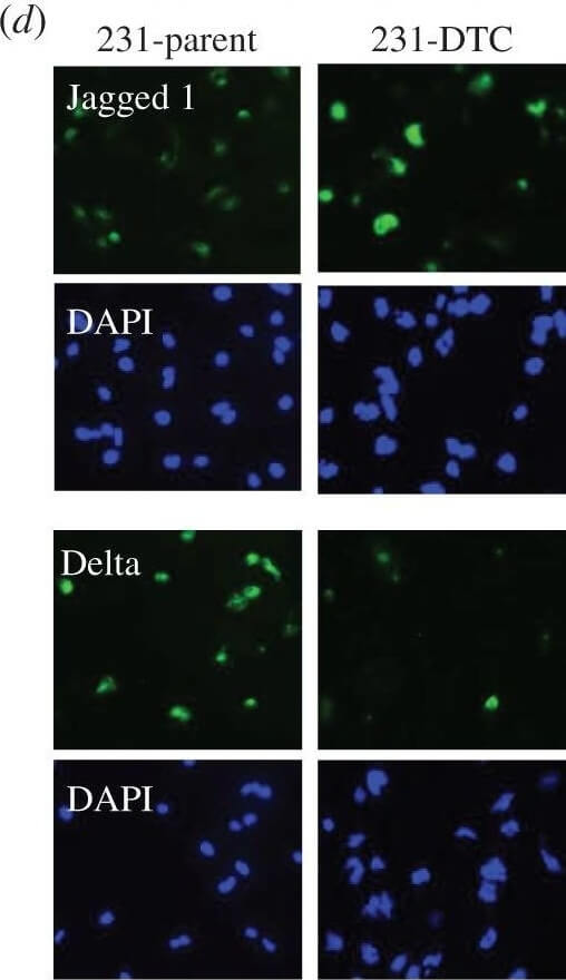
(a) Representative confocal microscopy shows CD44, CD24 and cleaved notch (NICD) in a population of drug naive MDA-MB-231. Yellow arrows indicate CD44HiCD24Lo (M) population of cells and the white arrows indicate the CD44HiCD24Hi (E/M) cells. Histogram (right panel) shows quantification of NICD in the distinct phenotype populations (M versus E/M). N = 3 biological replicates. (b) Schematic describes the experimental protocol to generate drug-tolerant cells (DTCs) parental MDA-MB-231 cells were treated with docetaxel at 100 nM (20* the IC50) and subsequently selected by substrate re-attachment and acute population outgrowth. (c) Representative confocal microscopy shows CD44, CD24 and NICD in the MDA-MB-231 parent and DTC populations. Right panel shows quantification of fluorescence intensity of each signal determined by at least 25 individual fields. N = 3 biological replicates. (d) Representative confocal microscopy shows Jagged and Delta expression in MDA-MB-231 parent and DTC. DAPI nuclear stain (blue). N = 3 biological replicates. Figure provided by CiteAb. Source: J R Soc Interface, PMID: 27170649.
* Mehrwertsteuer und Versandkosten nicht enthalten. Irrtümer und Preisänderungen vorbehalten