This protein A purified antibody was prepared from whole rabbit serum produced by repeated immunizations with a purified recombinant protein corresponding to full length SARS Coronavirus 3CL Protease. Lifesensors Inc. (www.lifesensors.com) prepared the 3CL Protease as follows: SUMO-3CL protease fusion was expressed in E. coli in LB medium and purified by Ni-NTA resin affinity chromatography (Qiagen). After the fusion was cleaved by the SUMO Protease (LifeSensors), the SUMO tag and protease were subtracted from the 3CL protease using MAC and the 3CL protease was finally purified using Anion Exchange Chromatography with the Macro-Prep High Q resin (BioRad) and size exclusion chromatography.
0.02 M Potassium Phosphate, 0.15 M Sodium Chloride, pH 7.2
Formulierung:
Lyophilized
Target-Kategorie:
SARS
Antibody Type:
Primary Antibody
Application Verdünnung:
ELISA: 1:10,000 - 1:50,000, IF Microscopy: User Optimized, WB: 1:2,000 - 1:10,000
Anwendungsbeschreibung:
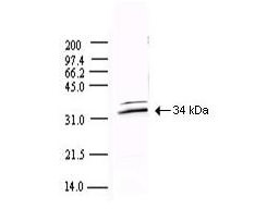
This protein A purified antibody has been tested for use in ELISA and by western blot. Specific conditions for reactivity should be optimized by the end user. Expect a band approximately 34 kDa in size corresponding to SARS 3CL Protease by western blotti
Western blot using Rocklands Protein A Purified anti-SARS CoV 3CL Protease antibody shows detection of a 34-kDa band corresponding to the protein. Approx. 100 ng of protein was loaded for SDS-PAGE and transferred onto nitrocellulose. The blot was incubated with a 1:5,000 dilution of the antibody at room temperature for 1 h followed by detection using IRDye(TM)800 labeled Goat-a-Rabbit IgG [H&L] (611-132-122) diluted 1:10,000. The fluorescence image was captured using the Odyssey Infrared Imaging System developed by LI-COR. IRDye is a trademark of LI-COR, Inc. Other detection systems will yield similar results.
* Mehrwertsteuer und Versandkosten nicht enthalten. Irrtümer und Preisänderungen vorbehalten