Anti-PID antibody was prepared from whole rabbit serum produced by repeated immunizations with a 17 amino acid peptide near the C-terminus of human PID.
Konjugation:
Unconjugated
Alternative Synonym:
PID Antibody, PID, MTA1L1, PID, Metastasis-associated protein MTA2, Metastasis-associated 1-like 1, MTA1-L1 protein
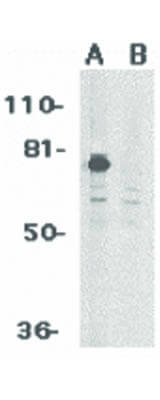
Anti-PID Antibody has been tested for use in ELISA, Western Blotting, Immunocytochemistry, Immunohistochemistry, and Immunofluorescence. Specific conditions for reactivity should be optimized by the end user. Expect a band at approximately 75 kDa in West
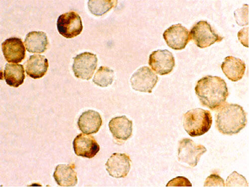
Immunocytochemistry of PID antibody. Cells: HeLa cells. Fixation: formalin fixed paraffin embedded. Antigen retrieval: not required. Primary antibody: PID antibody at 10 µg/mL for 1 h at RT. Secondary antibody: Peroxidase rabbit secondary antibody at 1:10,000 for 45 min at RT. Localization: PID is nuclear. Staining: PID brown nuclear stain.
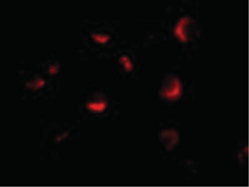
Immunofluorescence Microscopy of PID antibody. Tissue: HeLa cells. Fixation: 0.5% PFA. Antigen retrieval: not required. Primary antibody: PID antibody at 10 µg/mL for 1 h at RT. Secondary antibody: Fluorescein rabbit secondary antibody at 1:10,000 for 45 min at RT. Localization: PID is nuclear. Staining: PID as red fluorescent signal.
Western Blot of PID antibody. Lane 1: HeLa whole cell lysate. Lane 2: HeLa whole cell lysate in the presence of blocking peptide. Load: 35 µg per lane. Primary antibody: PID antibody at 1 µg/mL for overnight at 4C. Secondary antibody: Peroxidase rabbit secondary antibody at 1:10,000 for 45 min at RT. Block: 5% BLOTTO overnight at 4C. Predicted/Observed size: 75 kDa, 73 kDa for PID. Other band(s): PID splice variants and isoforms.
* Mehrwertsteuer und Versandkosten nicht enthalten. Irrtümer und Preisänderungen vorbehalten