0.02 M Potassium Phosphate, 0.15 M Sodium Chloride, pH 7.2
Formulierung:
Lyophilized
Target-Kategorie:
Human
Antibody Type:
Primary Antibody
Application Verdünnung:
ELISA: 1:10,000, WB: 1:1,000
Anwendungsbeschreibung:
This purified antibody has been tested in western blotting. Reactivity is also expected in ELISA, neutralizations, radioimmunoassay and immunohistochemistry. The endotoxin content is estimated to be <10 pg/µl by the LAL method. By western blot a band app
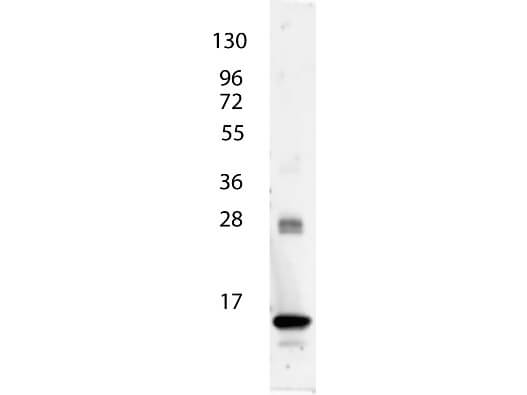
Rocklands anti-Human IL-3 antibody shows detection of a band ~15 kDa in size corresponding to recombinant human IL-3. The identity of the faint higher molecular weight band may represent a homodimer. Molecular weight markers are also shown (left). After transfer, the membrane was blocked overnight with 3% BSA in TBS followed by reaction with primary antibody at a 1:1,000 dilution. Detection occurred using peroxidase conjugated anti-Rabbit IgG (p/n 611-103-122) secondary antibody diluted 1:40,000 in blocking buffer (p/n MB-070) for 30 min at RT followed by reaction with FemtoMax(TM) chemiluminescent substrate. Image was captured using VersaDoc(TM) MP 4000 imaging system (Bio-Rad).
* Mehrwertsteuer und Versandkosten nicht enthalten. Irrtümer und Preisänderungen vorbehalten