Anti-IL-6 is an IgG fraction antibody prepared from rabbit antiserum after repeated immunizations with recombinant mouse IL-6 protein produced in E.coli.
0.02 M Potassium Phosphate, 0.15 M Sodium Chloride, pH 7.2
Formulierung:
Lyophilized
Target-Kategorie:
Mouse
Antibody Type:
Primary Antibody
Application Verdünnung:
ELISA: 1:10,000, IHC: User Optimized, WB: 1:1000
Anwendungsbeschreibung:
This purified antibody has been tested for use in Immunohistochemistry, western blotting, and flow cytometry in human and mouse cell lines. This product is suitable for ELISA. Specific conditions for reactivity should be optimized by the end user. Expect
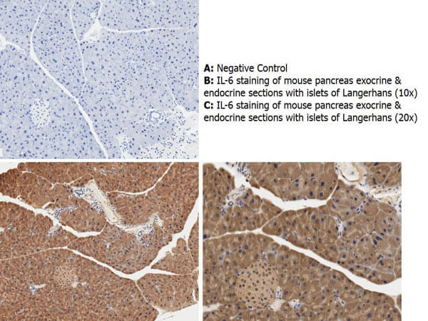
Immunohistochemistry with anti-IL-6 antibody showing cytoplasmic IL-6 staining in mouse pancreas exocrine and endocrine sections with islets of Langerhans at 10x and 20x (B & C). Staining was performed on Leica Bond system using the standard protocol. Formalin fixed/paraffin embedded tissue sections were subjected to antigen retrieval with E1 (Leica Microsystems) retrieval solution for 20 min and then incubated with rabbit anti-mouse IL-6 antibody at 1:50 dilution for 60 minutes. Biotinylated Anti-rabbit secondary antibody was used at 1:200 dilution to detect primary antibody. The reaction was developed using streptavidin-HRP conjugated compact polymer system and visualized with chromogen substrate, 33-diamino-benzidine substrate (DAB). The sections were then counterstained with hematoxylin to detect cell nuclei.
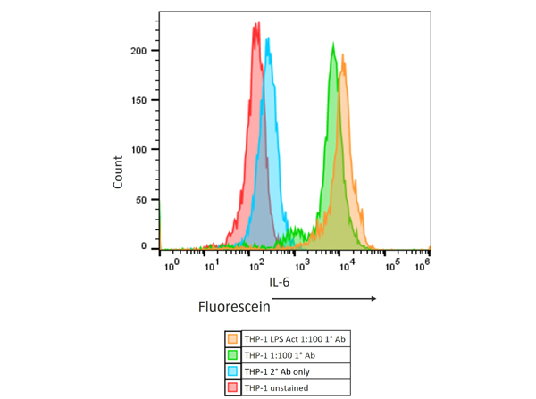
Flow Cytometry Results of Rabbit Anti-Mouse IL6 Antibody in human THP-1 cell line. The orange histogram represents the THP-1 cells that were activated with 100ng/mL LPS for 24 hours. The green histogram are untreated THP-1 cells. These two populations were intracellularly stained for 30 minutes at 4C in 1* BD Perm/Wash(TM) buffer. The primary stain was a 1:100 dilution of the Anti-Mouse IL-6 (RABBIT) Polyclonal Antibody (p/n 210-401-310, Lot 45055 [stock conc 1mg/mL]) and the secondary stain was the Anti-RABBIT IgG (H&L) (GOAT) Antibody Fluorescein Conjugated (p/n 611-1202 [1:400 dilution of 2mg/mL]). The secondary stain was for 30 minutes at 4C and was kept protected from light. The blue histogram is the THP-1 cells that were untreated and only stained with the secondary antibody. The red histogram is the untreated THP-1 cells that were not stained. Prior to staining, the cells for all conditions were permeabilized with BD Fixation/Permeabilization (TM) solution for 20 minutes at 4C. All washes and stains were performed in the BD 1* Perm/Wash(TM) buffer.
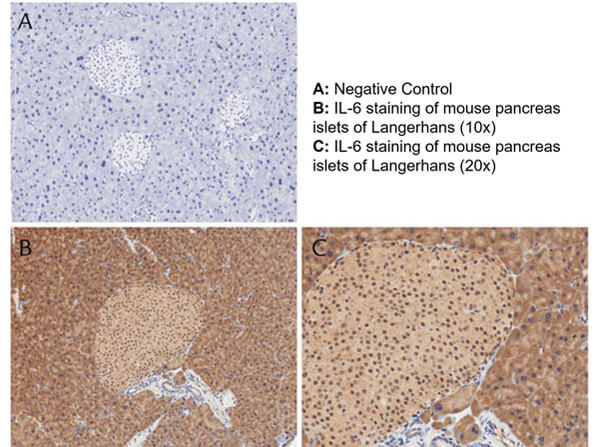
Immunohistochemistry with anti-IL-6 antibody showing positivity of islets of Langerhans (brown staining) and cytoplasmic staining in mouse pancreas at 10x and 20x (B & C). Staining was performed on Leica Bond system using the standard protocol. Formalin fixed/paraffin embedded tissue sections were subjected to antigen retrieval with E1 (Leica Microsystems) retrieval solution for 20 min and then incubated with rabbit anti-mouse IL-6 antibody at 1:50 dilution for 60 minutes. Biotinylated Anti-rabbit secondary antibody was used at 1:200 dilution to detect primary antibody. The reaction was developed using streptavidin-HRP conjugated compact polymer system and visualized with chromogen substrate, 33-diamino-benzidine substrate (DAB). The sections were then counterstained with hematoxylin to detect cell nuclei.
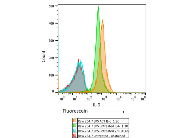
Flow Cytometry Results of Rabbit Anti-Mouse IL6 Antibody in mouse Raw 264.7 cell line. The orange histogram represents the Raw 264.7 murine cells that were activated with 100ng/mL LPS for 24 hours. The green histogram are untreated Raw 264.7 cells. These two populations were intracellularly stained for 30 minutes at 4C in 1* BD Perm/Wash(TM) buffer. The primary stain was a 1:30 dilution of the Anti-Mouse IL-6 (RABBIT) Polyclonal Antibody (p/n 210-401-310, Lot45055 [stock conc 1mg/mL]) and the secondary stain was the Anti-RABBIT IgG (H&L) (GOAT) Antibody Fluorescein Conjugated (p/n 611-1202 [1:400 dilution of 2mg/mL]). The secondary stain was for 30 minutes at 4C and was kept protected from light. The blue histogram is the Raw 264.7 murine cells that were untreated and only stained with the secondary antibody. The red histogram is the untreated Raw 264.7 murine cells that were not stained. Prior to staining, the cells for all conditions were permeabilized with BD Fixation/Permeabilization (TM) solution for 20 minutes at 4C. All washes and stains were performed in the BD 1* Perm/Wash(TM) buffer.
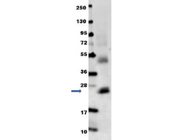
Anti-mouse IL-6 antibody in western blot shows detection of recombinant mouse IL-6 raised in E.coli. Recombinant truncated protein (0.1 µg, 21.7 kDa) was loaded on to an SDS-PAGE
* Mehrwertsteuer und Versandkosten nicht enthalten. Irrtümer und Preisänderungen vorbehalten