apoLipoprotein Type B was isolated from human plasma by density gradient centrifugation followed by HPLC purification.
Konjugation:
Unconjugated
Alternative Synonym:
goat anti-Apolipoprotein B antibody, Apo B-100, Apo B-48, APOB protein antibody, Apolipoprotein-B 100 antibody, Apolipoprotein B 48 antibody, Apolipoprotein B antibody, FLDB antibody
0.125 M Sodium Borate, 0.075 M Sodium Chloride, 0.005 M EDTA, pH 8.0
Formulierung:
Liquid (sterile filtered)
Target-Kategorie:
Human
Antibody Type:
Primary Antibody
Application Verdünnung:
ELISA: 1:2,000 - 1:10,000, IHC: 1:50 - 1:500, IF Microscopy: User Optimized, IP: 1:100, WB: 1:200 - 1:1,000
Anwendungsbeschreibung:
Anti-apoLipoprotein antibodies have been used for indirect trapping ELISA for quantitation of antigen in serum using a standard curve, for immunoprecipitation and for western blotting for highly sensitive qualitative analysis.
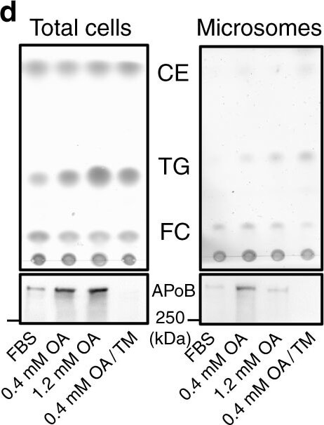
LDs are present in the lumen of the type I NR. a Huh7 treated with OA/TM for 24-48h harbored LDs in the lumen of the ER (left, arrowheads) and within the type I NR (right, arrows mark the NR). INM: inner nuclear membrane, ONM: outer nuclear membrane. Bars, 0.2µm. b Huh7-expressing HRP-KDEL treated with OA/TM for 24h. DAB precipitated in the type I NR lumen (arrows). Bar, 0.5µm. c Mouse hepatocytes in vivo after high-fat diet feeding for 6 weeks and TM injection. Lumenal LDs were observed in the ER (arrowheads), the nuclear cistern (arrowhead in the inset) (left figure, Bar, 0.5µm), and in the type I NR (arrows mark the NR) (right figure, Bar, 0.2µm). They contained more nuclear LDs than the control fed the high-fat diet and injected with vehicle alone. MeanSD of three independent experiments. *p<0.01, Students t test. d Microsomes of Huh7 treated with none, 0.4mm OA, 1.2mm OA, or OA/TM for 48h. The OA/TM-treated cell microsome contained triglycerides (TG) and cholesterol esters (CE) most abundantly (by thin layer chromatography), but showed the lowest amount of ApoB (by Western blotting). e Three different kinds of LDs in the nuclear area: Nucleoplasmic LDs (A), NR-lumenal LDs (within the type I NR) (B), and cytoplasmic LDs (within the type II NR) (C). f Nucleoplasmic LDs (arrowheads) and NR-lumenal LDs (arrows) are distinguished by whether they are outside of or within LBR rings, respectively. Huh7 treated with OA/TM for 48h. Both LDs were reduced by MTPi (100nm BAY 13-9952). Bar, 10µm. See also Supplementary Fig.2e. g The number of nucleoplasmic LDs, NR-lumenal LDs, and cytoplasmic LDs within the type II NR were counted in randomly taken electron micrographs of Huh7 treated with OA/TM for 48-72h. Box plot of pooled data from three independent experiments. The average is shown by+. Number of nuclei examined=128.Source data are provided as aSource data file Figure provided by CiteAb. Source: Nat Commun, PMID: 30692541.
* Mehrwertsteuer und Versandkosten nicht enthalten. Irrtümer und Preisänderungen vorbehalten