Anti-apoLipoprotein antibodies have been used for indirect trapping ELISA for quantitation of antigen in serum using a standard curve, for immunoprecipitation and for western blotting for highly sensitive qualitative analysis.
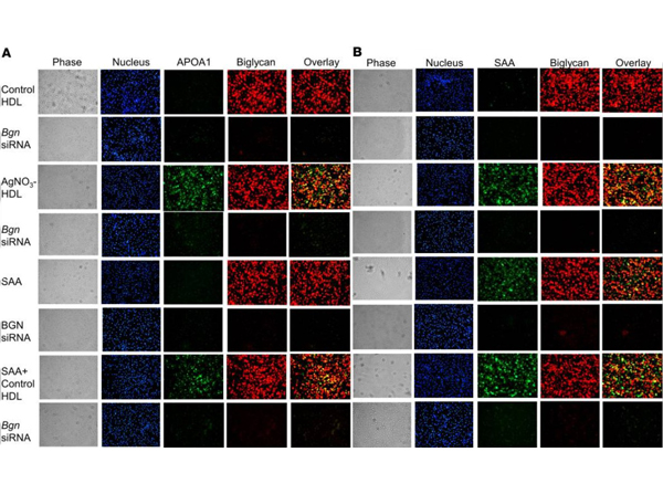
Immunofluorescence of Anti-Apolipoprotein AI.HDL isolated from AgNO3-injected mice colocalizes with biglycan at the cell surface of peritoneal macrophages. HDL from PBS- and AgNO3-injected C57BL/6 mice was isolated. After exposure to these HDL preparations (50 µg protein/mL) for 6 hours, TG-elicited peritoneal macrophages fromSaa3-/-were fixed in 2% formalin for 5 minutes (AandB). After extensive washing, (A) APOA1 and biglycan were stained using anti-biglycan (red) and anti-APOA1 (green) antibodies, or (B) SAA and biglycan were stained using anti-biglycan (red) and ant-SAA (green) antibodies and photographed by fluorescence microscopy (Nikon Eclipse 80i, original magnification, *200). Figure 5. PMID: 32970631.
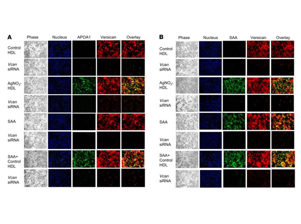
Immunofluorescence of Anti-Apolipoprotein AI.Versican colocalizes with HDL isolated from AgNO3-injected mice at the cell surface of 3T3-L1 adipocytes. Free SAA and HDL from PBS- or AgNO3-injected C57BL/6 mice were isolated. Some adipocytes were transfected with siRNA specific for versican (Vcan) for 3 days. After exposure to free SAA (15 µg/mL) and/or these HDL preparations (50 µg protein/mL) for 6 hours, 3T3-L1 adipocytes were fixed in 2% formalin for 5 minutes. After extensively washing, (A) APOA1 and versican were stained using anti-versican (shown in red) and anti-APOA1 (shown in green) antibodies, or (B) SAA and versican were stained using anti-versican (shown in red) and anti-SAA (shown in green) antibodies and photographed by fluorescence microscopy (Nikon Eclipse 80i, original magnification, *200). Cell nuclei were counterstained with DAPI (blue). Cell morphology was shown by phase-contrast photography (left). Merged fluorescence (overlay) is shown in yellow. Figure 2. PMID: 32970631.
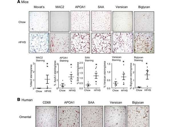
Immunohistochemistry of Anti-Apolipoprotein AI.Immunohistochemical staining of versican and biglycan shows a similar distribution with the staining of SAA and APOA1 in the epididymal white adipose tissue from mice fed an HFHS diet and in the omental fat tissue from human obese subjects undergoing gastric bypass surgery. (A) Mice were fed a chow or HFHS diet for 16 weeks, after which adipose tissue was obtained and immunostained with the antibodies shown. Tissues were photographed using microscopy (original magnification, *60). Quantitation of the immunostaining is shown in the lower panel (n= 5-7, mean SEM). *P< 0.001 vs. chow. (B) Omental fat was obtained from gastric bypass patients and immunostained with the antibodies shown. Tissues were photographed using microscopy (original magnification, *60). The pictures of MAC2, CD68, versican, and biglycan staining are from our previous publication (17), APOA1 and SAA staining were performed on adjacent sections. Figure 1. PMID: 32970631.
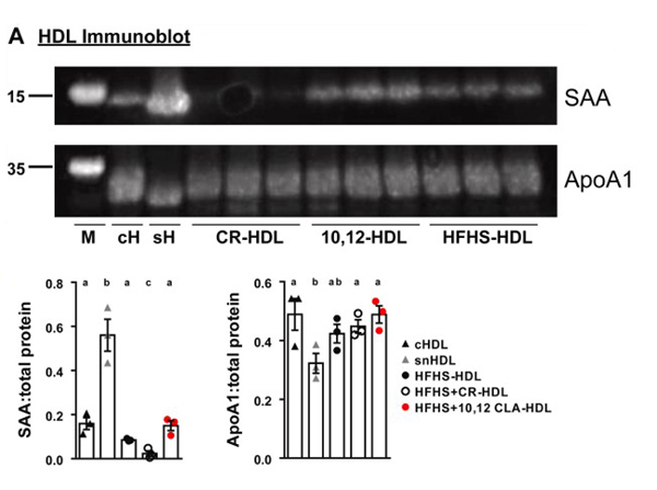
Western Blot of Anti-Apolipoprotein AI.Proteomics confirmation and hepatic gene expression. A: HDL preparations from the indicated treatment groups were subjected to immunoblot and probed for Saa (marker= 15 kD) and Apoa1 (marker= 35 kD). Densitometry was performed using ImageJ software. Control HDL samples were pooled,n= 3 for treatment groups. Different letters indicate significant differences, assessed using one-way ANOVA with multiple comparisons (Tukey) (P< 0.05). cH, control HDL from mice injected with saline, CR-HDL, HDL from mice fed an HFHS diet that were calorically restricted, 10,12-HDL, HDL from mice fed an HFHS diet containing 10,12 CLA, HFHS-HDL, HDL from mice fed an HFHS diet, sH, HDL from mice injected with silver nitrate. B: Liver gene expression was quantified from the indicated treatment groups.n= 8 mice/group, *P< 0.05 from HFHS. Fig.5. PMID: 35714730.
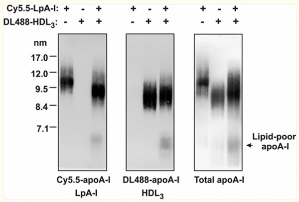
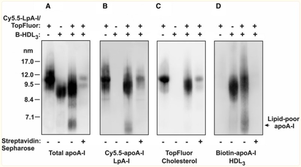
Western Blot of Anti-Apolipoprotein AI.Lipoprotein particle remodeling is induced by the interaction of nascent LpA-I (apo AI [apolipoprotein AI] containing particles formed by incubating ABCA1 [ATP-binding cassette transporter 1]-expressing cells with apo AI) and HDL3in vitro.Fluorescently labeled LpA-I (Cy5.5-LpA-I) and HDL3(DL488-HDL3) were incubated f
* Mehrwertsteuer und Versandkosten nicht enthalten. Irrtümer und Preisänderungen vorbehalten