This affinity purified antibody was prepared from whole goat serum produced by repeated immunizations with a synthetic peptide corresponding to the N-Terminal region near aa 1-25 of Human AP2A1 and AP2A2 proteins.
Konjugation:
Unconjugated
Alternative Synonym:
goat anti-AP2A Antibody, AP-2 complex subunit alpha-1, Adapter-related protein complex 2 alpha-1 subunit, Adaptor protein complex AP-2 subunit alpha-1 Alpha1-adaptin, Plasma membrane adaptor HA2/AP2, Clathrin assembly protein complex 2 alpha-A large chain, AP2A2, ADTAB, CLAPA2, HIP9, HYPJ, KIAA0899
This affinity purified antibody has been tested for use in ELISA and by western blot. Specific conditions for reactivity should be optimized by the end user. Expect a band approximately 104-107 kDa in size corresponding to AP2A1 or AP2A2 proteins by west
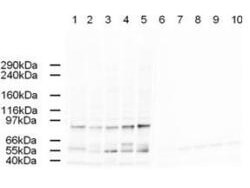
Western blot using Rocklands Affinity Purified anti-AP2A antibody shows detection of a band just below 100 kDa corresponding to Human AP2A1. Lane 1: HeLa nuclear extract (p/n W09-001-367), Lane 2: HeLa (p/n W09-000-364), Lane 3: 293 (W09-000-365), Lane 4: A431 (p/n W09-000-361), and Lane 5: Jurkat whole cell lysate (p/n W09-001-370). In lanes 6-10 the antibody was preincubated with 1 µg/ml of the immunizing peptide which effectively blocks the specific reactivity of this antibody with AP2A. Approximately 20 µg of each lysate was run on a SDS-PAGE and transferred onto nitrocellulose followed by reaction with a 1:500 dilution of anti-AP2A antibody. Detection occurred using a 1:5,000 dilution of HRP-labeled Rabbit anti-Goat IgG (p/n 605-4302) for 1 hour at room temperature. A chemiluminescence system was used for signal detection (Roche) using a 60-sec exposure time.
* Mehrwertsteuer und Versandkosten nicht enthalten. Irrtümer und Preisänderungen vorbehalten