Anti-Brn3b antibody was prepared from whole goat serum produced by repeated immunizations with a synthetic peptide corresponding to a near N-terminal portion of mouse Pou4f2 conjugated to Keyhole Limpet Hemocyanin (KLH).
Konjugation:
Unconjugated
Alternative Synonym:
Goat Anti-Brn3b Antibody, Goat Anti-Pou4f2 Antibody, POU domain, class 4, transcription factor 2, Brain-specific homeobox/POU domain protein 3B, Brn-3B, Brain-3B, Brn-3.2
Anti-Brn3b Antibody has been tested in WB, IHC, and IF. Expect a band at ~43kDa in western blot using appropriate lysates. Positive controls used: Rat pup PND2-6 - minimal cortex brain lysate in WB, Mouse testis in IHC, Post-natal rat pup (PND1) heteroge
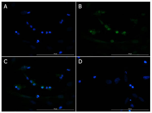
Immunofluorescence of Goat Anti-Brn3b Antibody. Cell Line: Post-natal rat pup (PND1) heterogeneous brain cells. Fixative: 4% PFA. Permeabilization: 0.3% Triton X-100. Primary Antibody: Anti-Brn3b at 5µg/mL overnight at 2-8C. Secondary Antibody: Donkey Anti-Goat IgG DyLight(TM)488 (p/n 605-741-125) at 15µg/mL for 1 hr at RT. Nuclear Counterstain: DAPI. Staining: (A). DAPI. (B). Brn3b + secondary. (C). Merge A+B. (D). secondary only. Expected localization: Nuclear, nuclear speckle.
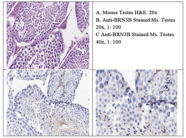
Immunohistochemistry of Goat Anti-Brn3b Antibody. Tissue: Mouse Testis. Fixative: none. Antigen Retrieval: HIER using Citrate Buffer for 20 minutes. Primary Antibody: Anti-Brn3b at 1:100 for 30 minutes at RT. Secondary Antibody: Donkey Anti-Goat IgG HRP 4µl/mL for 20 minutes at RT. Counterstain: Hematoxylin. Substrate: DAB. Analysis: BRN3B staining of human testes shows intense punctate intranuclear staining of Sertoli cell of the seminiferous tubules which is the expected staining pattern.
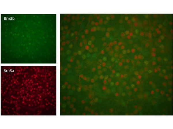
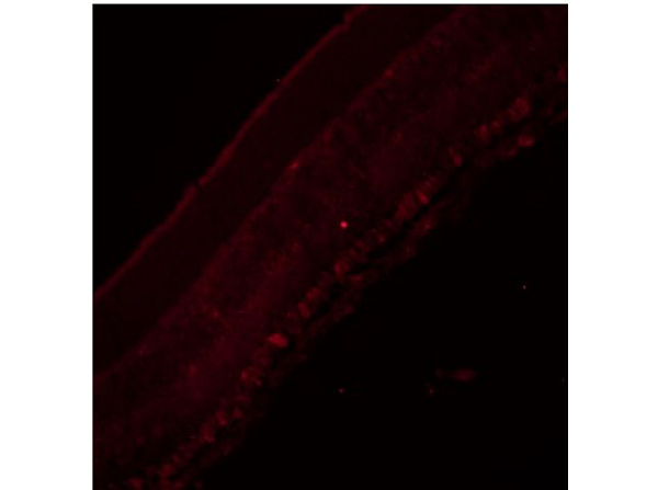
Immunofluorescence of Goat Anti-Brn3b Antibody. Tissue: Mouse Retina Tissue. Permeabilization: 0.5%Triton X-100 per 45mins. Primary Antibody: Anti-Brn3b at 1:200 for 3 days at 2-8C. Incubate secondary antibody for 2hrs. Incubate HOECSHT for 30 mins. Fluoro-G mounting. Courtesy of Viviane Valenca at University of Utah.
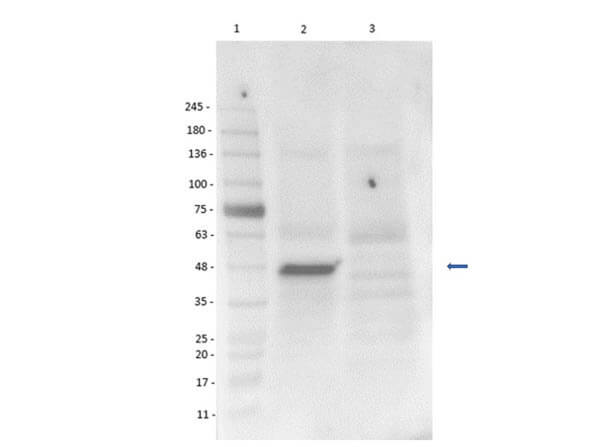
Western Blot of Goat Anti-Brn3b Antibody. Lane 1: Opal Prestained Molecular weight ladder (p/n MB-210-0500). Lane 2: Rat pup PND2-6 - minimal cortex brain lysate (35µg) [+]. Lane 3: HEK293T whole cell lysate (35µg) [-]. Primary Antibody: Anti-Brn3b at 1:1000 overnight at 2-8C. Secondary Antibody: Donkey anti-Goat IgG HRP (p/n 605-703-125) at 1:40,000 for 30 minutes at RT. Block: BlockOut Buffer (p/n MB-073). Exposure: 12 sec. Predicted MW: ~43kDa (rat), ~35, ~43kDa (mouse). Observed MW: ~47kDa.
Immunofluorescence of Goat Anti-Brn3b Antibody. Tissue: Mouse Retina Tissue. Retrieval: 10mM Sodium Citrate pH6 retrieval and 10mM Tris-base 1mM EDTA 0.05% Tween 20 pH9. Permeabilization: 0.5% Triton X-100 per 15 mins. Primary Antibody: Anti-Brn3b at 1:200 overnight at 2-8C. Incubate secondary antibody 2 hrs. Incubate HOECSHT 10 mins. Fluoro-G mounting. This antibody works with or without retrieval. Courtesy of Viviane Valenca at University of Utah.
* Mehrwertsteuer und Versandkosten nicht enthalten. Irrtümer und Preisänderungen vorbehalten