The immunogen is a Green Fluorescent Protein (GFP) fusion protein corresponding to the full length amino acid sequence (246aa) derived from the jellyfish Aequorea victoria.
Konjugation:
DL488
Alternative Synonym:
goat anti-GFP Antibody DyLight(TM) 488 Conjugation, DyLight(TM) 488 conjugated goat anti-GFP antibody, Green Fluorescent Protein, GFP antibody, Green Fluorescent Protein antibody, EGFP, enhanced Green Fluorescent Protein, Aequorea victoria, Jellyfish
Anti-GFP Dylight(TM) 488 Conjugated Antibody has been tested by dot blot and western blot. The emission spectra for this DyLight(TM) conjugate match the principle output wavelengths of most common fluorescence instrumentation.
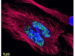
Multi-color Immunofluorescence Microscopy using DyLight(TM) dyes. Tissue: human breast carcinoma. Fixation: 0.5% PFA. Antigen retrieval: not required. Primary antibody: Anti-Histone and Anti-Tubulin antibody at 10 µg/mL for 1 h at RT. Secondary antibody: DyLight(TM) 488 conjugate and DyLight(TM) 549 conjugate goat secondary antibody at 1:10,000 for 45 min at RT. Localization: Histone is nuclear and Tubulin is cytoplasmic. Staining: Anti-Histone detection using a DyLight(TM) 488 conjugate (green) fluorescent signal and Anti-Tubulin was detected using a DyLight(TM) 549 conjugate (red) fluorescent signal. Nuclei were counter-stained using DAPI (blue).
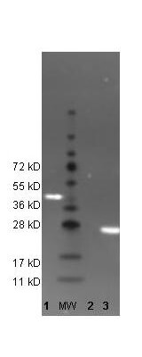
Western Blot of DyLight(TM) 488 conjugated anti-GFP antibody to detect GFP control proteins. Lane 1: His-Sumo-GFP. Lane: Molecular Weight. Lane 2: Beta-Galactosidase (negative control). Lane 3: recombinant GFP control protein (000-001-215). Load: 35 µg per lane. Primary antibody: none. Secondary antibody: DyLight(TM) 488 conjugated anti-GFP goat secondary antibody at 1:5,000. Block: MB-070 for 2 hr at RT. Predicted/Observed size: 27kDa/54kDa, 27kDa for rGFP/~45kDa His-Sumo-GFP. Other band(s): none.
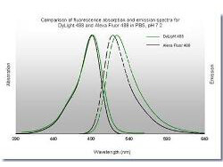
Comparison of fluorescence absorbtion and emission spectra for DyLight(TM) 488 and Alexa Fluor 488 in PBS, pH7.2. The emission spectra for this DyLight(TM) conjugate match the principle output wavelengths of most common fluorescence instrumentation.
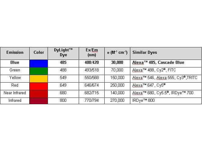
Properties of DyLight(TM) Fluorescent Dyes. The emission spectra for this DyLight(TM) conjugate match the principle output wavelengths of most common fluorescence instrumentation.
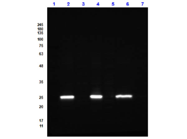
Western Bot of Goat Anti-GFP (GOAT) Antibody DyLight(TM) 488 Conjugated. Lane 1: Opal Prestained Molecular Weight Marker (p/n MB-210-0500). Lane 2: GFP / HEK293T Whole Cell Lysate Reduced [0.05/10µg]. Lane 3: HEK293T Whole Cell Lysate Reduced (p/n W09-001-GX5) [10µg]. Lane 4: GFP / NIH-3T3 Whole Cell Lysate Reduced [0.05/10µg]. Lane 5: NIH-3T3 Whole Cell Lysate Reduced (p/n W10-000-358) [10µg]. Lane 6: GFP / PC-12 Whole Cell Lysate Reduced [0.05/10µg].Lane 7: PC-12 Whole Cell Lysate Reduced (p/n W12-001-GL9) [10µg]. Secondary Antibody: Goat Anti-GFP (GOAT) Antibody DyLight(TM) 488 Conjugated at 1.0µg/mL overnight at 2-8C. Blocking Buffer: Blocking Buffer for Fluorescent Western Blotting (p/n MB-070) for 1 hour at RT. Expect: 27kDa.
* Mehrwertsteuer und Versandkosten nicht enthalten. Irrtümer und Preisänderungen vorbehalten