The immunogen is a Green Fluorescent Protein (GFP) fusion protein corresponding to the full length amino acid sequence (246aa) derived from the jellyfish Aequorea victoria.
Konjugation:
Unconjugated
Alternative Synonym:
rabbit anti-GFP antibody, Green Fluorescent Protein, GFP antibody, Green Fluorescent Protein antibody, EGFP, enhanced Green Fluorescent Protein, Aequorea victoria, Jellyfish
0.02 M Potassium Phosphate, 0.15 M Sodium Chloride, pH 7.2
Formulierung:
Liquid (sterile filtered)
Antibody Type:
Primary Antibody
Application Verdünnung:
ELISA: 1:20,000 - 1:120,000, Flow Cytometry: User Optimized, IHC: 1:200 - 1:3,000, IF Microscopy: 1:500 - 1:5,000, IP: User Optimized, WB: 1:500 - 1:5,000
Anwendungsbeschreibung:
Anti-GFP antibody is designed to detect GFP and its variants. GFP antibody has been tested by western blot and ELISA. This product can be used to detect GFP by ELISA (sandwich or capture) for the direct binding of antigen and recognizes wild type, recomb
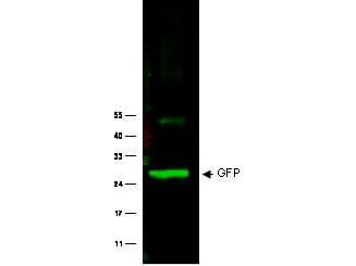
Western Blot of Rabbit anti-GFP antibody. Lane 1: Wild type GFP (0.1 µg) was used to spike HeLa whole cell lysate. Lane 2: none. Load: 30 µg per lane. Primary antibody: GFP antibody at 1:1000 for overnight at 4C. Secondary antibody: IRDye800(TM) Goat-a-Rabbit IgG [H&L] MX10 (611-132-122) at 1:10,000 for 45 min at RT. Block: 5% BLOTTO in PBS overnight at 4C. Predicted/Observed size: 27 kDa for epitope tag GFP. Other band(s): none.
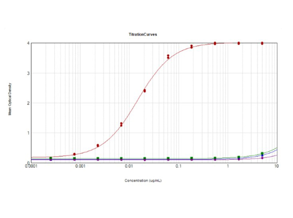
ELISA results of purified Rabbit Anti-GFP Antibody. Each well was coated in 10 µg of antigen GFP [Red Line], human IgG [Green Line], Mouse IgG [Blue Line], and Rat IgG [Purple Line]. The starting dilution of antibody was 5 µg/mL and the X-axis represents the Log10 of a 3-fold dilution. The titer is 1:67,700. This titration is a 4-parameter curve fit where the IC50 is defined as the titer of the antibody. Assay performed using 1% Fish Gel, TMB Substrate (p/n TMB-1000), and Goat Anti-Rabbit IgG Antibody HRP (p/n 611-103-122).
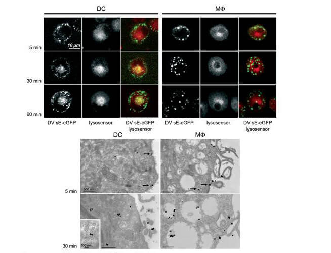
Immuno-microscopy of Rabbit anti-GFP antibody. Monocyte derived dendritic cells and dermal macrophages were challenged and directly visualized with eGFP labeled Dengue virus to localize sequestration of virus particles in the different cells (upper). The location of the GFP was confirmed by TEM (lower magnified view) using Rockland rabbit anti GFP Primary antibody (1:200) and a gold labeled secondary antibody. As referenced in: Kwan W-H, Navarro-Sanchez E, Dumortier H, Decossas M, Vachon H, et al. (2008) Dermal-Type Macrophages Expressing CD209/DC-SIGN Show Inherent Resistance to Dengue Virus Growth. PLoS Negl Trop Dis 2(10): e311. doi:10.1371/journal.pntd.0000311
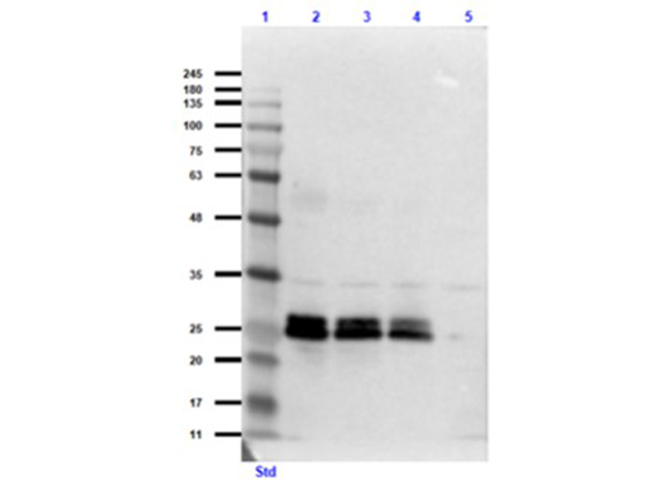
Western Blot of Rabbit Anti-GFP Antibody. Lane 1: Opal Prestained Molecular Weight Ladder (p/n MB-210-0500). Lane 2: GFP (p/n 000-001-215) / HeLa Lysate (p/n W09-000-364) [0.1µg/10.0µg]. Lane 3: GFP (p/n 000-001-215) / HeLa Lysate (p/n W09-000-364) [0.05µg/10.0µg]. Lane 4: GFP (p/n 000-001-215) / HeLa Lysate (p/n W09-000-364) [0.03µg/10.0µg]. Lane 5: HeLa Lysate (p/n W09-000-364) [10.0µg]. Primary Antibody: Rabbit Anti-GFP Antibody at 1.0µg/mL overnight at 2-8C. Secondary Antibody: Goat Anti-Rabbit IgG MX 10 Peroxidase (p/n 611-103-122) at 1:70,000 for 30mins at RT. Block: Blocking Buffer for Fluorescent Western Blotting (p/n MB-070) for 60 mins at RT. Expect: ~27kDa.
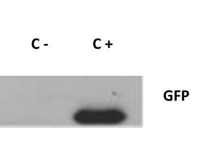
Western Blot of Rabbit anti-GFP antibody. Lane 1: 293FT cells transfected with CDK4 dominant negative (C-). Lane 2: 293FT cells poitive control (C+). Load: 25 µg per lane. Primary antibody: GFP antibody at 1:400 for overnight at 4C. Secondary antibody: IRDye800(TM) rabbit secondary antibody at 1:10,000 for 45 min at RT. Block: 5% BLOTTO overnight at 4C. Predicted/Observed size: 27 kDa for GFP.
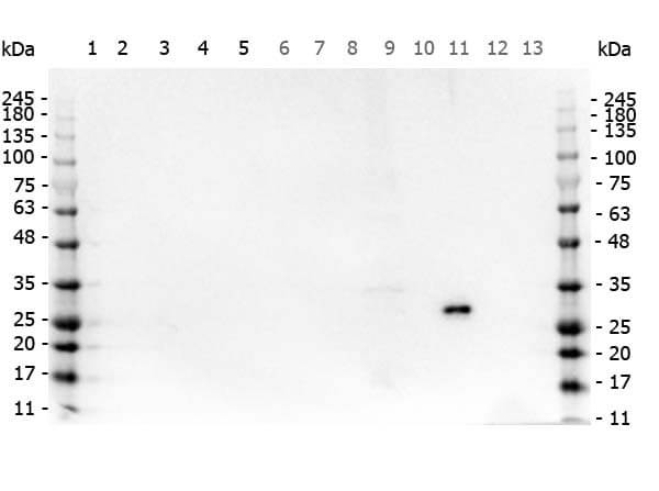
Western Blot of Rabbit anti-GFP antibody. Marker: Opal Pre-stained ladder (p/n MB-210-0500). Lane 1: HEK293 lysate (p/n W09-000-365). Lane 2: HeLa Lysate (p/n W09-000-364). Lane 3: CHO/K1 Lysate (p/n W07-000-357). Lane 4: MDA-MB-231 (p/n W09-001-GK6). Lane 5: A431 Lysate (p/n W09-000-361). Lane 6: Jurkat Lysate (p/n W09-001-370). Lane 7: NIH/3T3 Lysate (p/n W10-000-358). Lane 8: E-coli HCP Control (p/n 000-001-J08). Lane 9: FLAG Positive Control Lysate (p/n W00-001-383). Lane 10: Red Fluorescent Protein (p/n 000-001-379). Lane 11: Green Fluorescent Protein (p/n 000-001-215). Lane 12: Glutathione-S-Transferase Protein (p/n 000-001-200). Lane 13: Maltose Binding Protein (p/n 000-001-385). Load: 10 µg of lysate or 50ng of purified protein per lane. Primary antibody: GFP antibody at 1ug/mL overnight at 4C. Secondary antibody: Peroxidase rabbit secondary antibody (p/n 611-103-122) at 1:30,000 for 60 min at RT. Blocking Buffer: 1% Casein-TTBS (p/n MB-082) for 30 min at RT. Predicted/Observed size: 30 kDa for GFP.
* Mehrwertsteuer und Versandkosten nicht enthalten. Irrtümer und Preisänderungen vorbehalten