This antibody was affinity purified from whole rabbit serum prepared by repeated immunizations with a synthetic peptide corresponding to an internal region of rabbit MAPKAP Kinase 2 conjugated to KLH using maleimide. A terminal cysteine residue was added to facilitate coupling.
Konjugation:
Unconjugated
Alternative Synonym:
rabbit anti-MAPKAP kinase 2 antibody, MAP kinase-activated protein kinase 2, MAPK-activated protein kinase 2, MAPKAP kinase-2, MK2, mitogen-activated protein kinase-activated protein kinase-2, MAPKAP-K2, MAPKAPK-2, MK-2, MK2
0.02 M Potassium Phosphate, 0.15 M Sodium Chloride, pH 7.2
Formulierung:
Liquid (sterile filtered)
Target-Kategorie:
Human
Antibody Type:
Primary Antibody
Application Verdünnung:
ELISA: 1:15,000, IHC: User Optimized, IP: User Optimized, WB: 1:2,000
Anwendungsbeschreibung:
Anti-MAPKAP kinase 2 antibody is tested by western blotting and suitable for ELISA, immunoprecipitation and kinase assays. Although not tested, this antibody is likely functional in immunohistochemistry and other immunological methods. The antibody detec
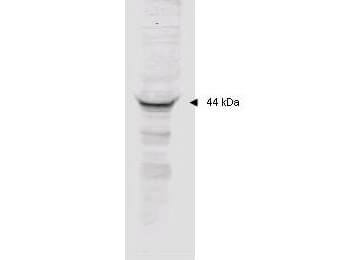
Affinity purified anti-MAPKAP Kinase 2 polyclonal antibody detects MK2 in unstimulated human HeLa whole cell lysate by western blot. Polyclonal rabbit-anti-MAPKAP Kinase 2 used at a 1:2000 dilution to detect 20 ug of whole cell lysate containing MK2. This antibody detects a single 44 kDa protein as indicated in crude extracts prepared from unstimulated human HeLa cell lysates. A 4-20% gradient gel was used to separate the protein by SDS-PAGE. The protein was transferred to nitrocellulose using standard methods. After blocking the membrane was probed with the primary antibody for 1 h at room temperature followed by washes and reaction with a 1:5,000 dilution of IRDye(TM)800 conjugated Gt-a-Rabbit IgG [H&L] (code 611-132-122) for 30 min at room temperature. LICORs Odyssey Infrared Imaging System was used to scan and process the image. Other detection systems will yield similar results.
* Mehrwertsteuer und Versandkosten nicht enthalten. Irrtümer und Preisänderungen vorbehalten