This antibody was purified from whole rabbit serum prepared by repeated immunizations with V5 epitope tag peptide corresponding to aa 95-108 of the V protein conjugated to KLH using maleimide.
Konjugation:
Unconjugated
Alternative Synonym:
Rabbit Anti-V5 Epitope Tag Antibody, Rabbit Anti V5 Epitope Tag Antibody, Rabbit Anti-V5 Tag
Anti-V5 is optimally suited for monitoring expression of V5-tagged fusion proteins. The V5 epitope tag is derived from a small epitope (Pk) present on the P and V proteins of the paramyxovirus of simian virus 5 (SV5). The V5 tag is usually used with all
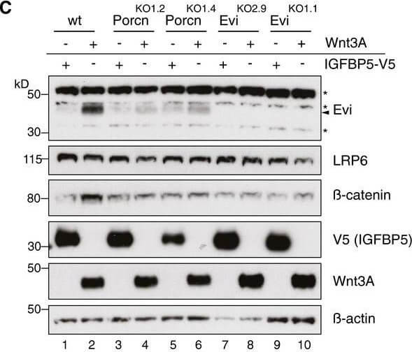
Evi stabilization is dependent on Wnt palmitoylation. Western blot analysis of endogenous Evi in wt, PorcnKO, or EviKO HEK293T cells upon overexpression of Wnt3A or IGFBP5-V5. PorcnKO1.2 and PorcnKO1.4 indicate clone 2 and clone 4 of PorcnKO HEK293T cells generated with Porcn sgRNA1 (Appendix Fig S3). Clonal EviKO HEK293T cells were generated with Evi sgRNA2 (EviKO2.9, clone 9) or Evi sgRNA1 (EviKO1.1, clone 1, Appendix Fig S2). Increase in total beta-catenin protein served as control for Wnt pathway activation. Figure 2C. Source: EMBO J, PMID: 29378775.
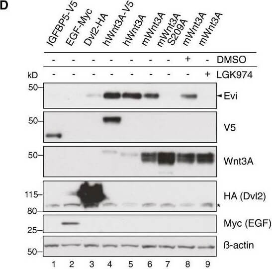
Evi stabilization is dependent on Wnt palmitoylation. Western blot analysis of endogenous Evi in HEK293T cells transfected with the indicated overexpression plasmids. When indicated, the cells were additionally treated with 5 µM LGK974 for 48 h. Figure 2D. Source: EMBO J, PMID: 29378775.
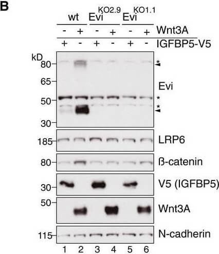
Wnt ligand production increases Evi protein levels. Wild-type (wt) or EviKO HEK293T cells were transfected with Wnt3A or IGFBP5-V5 expression plasmids and subjected to Western blot analysis. Specific Evi bands are indicated by arrows and unspecific bands by asterisks. Endogenous Evi is not only detectable as a monomeric form (46 kDa) but also as SDS-resistant dimers (80 kDa). Clonal EviKO HEK293T cells were generated via CRISPR/Cas9 using Evi sgRNA 2 (EviKO2.9) or Evi sgRNA 1 (EviKO1.1, Appendix Fig S2). Figure 1B. Source: EMBO J, PMID: 29378775.
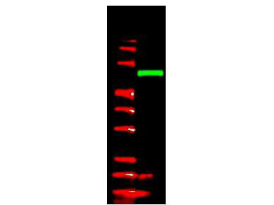
Anti-V5 epitope tag polyclonal antibody detects V5-tagged recombinant protein by western blot. This antibody was used at 1.0 µg/ml to detect 0.05 µg (lane 2) of full-length recombinant mouse serum albumin containing the V5 epitope tag at the carboxy end. Comparison to MW markers (lane 1) indicates detection of monomeric V5 tagged albumin. A 4-20% gradient gel was used to separate the protein by SDS-PAGE under non-reducing conditions. The protein was transferred to nitrocellulose using standard methods. After blocking the membrane was probed with the primary antibody overnight at 4 C followed by washes and reaction with a 1:10,000 dilution of IRDye 800 conjugated Gt-a-Rabbit IgG [H&L] (code 611-132-122) for 45 min at room temperature. LICORs Odyssey Infrared Imaging System was used to scan and process the image. Other detection systems will yield similar results.
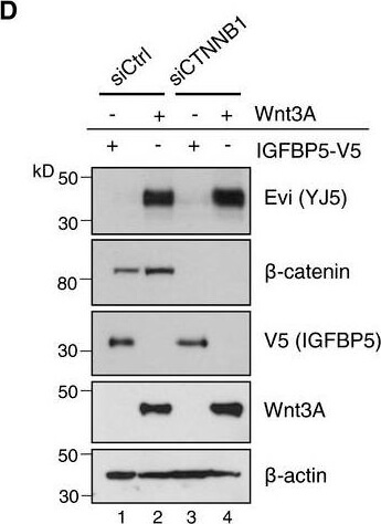
Evi is not transcriptionally regulated by Wnt. D'Twenty-four hours after reverse transfection with Ctrl or CTNNB1 siRNA, HEK293T cells were transfected with Wnt3A or IGFBP5-V5 expression plasmids and analyzed (D) for the indicated proteins via immunoblotting or (D') for canonical Wnt activity using the TCF-Luciferase Wnt reporter assay. Immunoblotting is representative of three independent experiments, and Wnt reporter activity was calculated as mean from three independent experiments s.d. Figure EV1D. Source: EMBO J, PMID: 29378775.
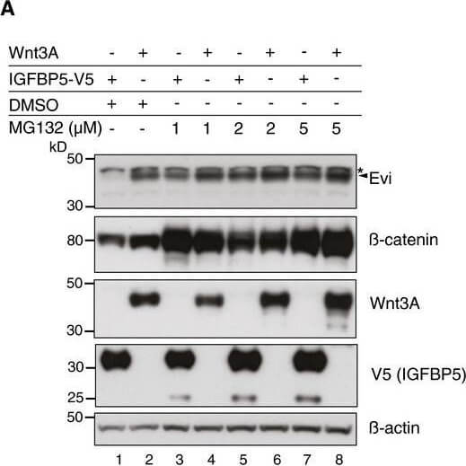
Evi is ubiquitinated and degraded from the ER by the proteasome. HEK293T cells were transfected with Wnt3A or IGFBP5-V5 expression plasmids and treated for 24 h with DMSO, MG132, or bortezomib in the indicated concentrations. Figure EV3B. Source: EMBO J, PMID: 29378775.
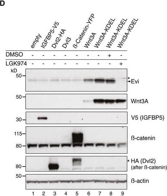
Evi poly-ubiquitination is regulated by the presence of Wnt proteins. HEK293T cells were transfected with the indicated plasmids and additionally treated with 5 µM LGK974 for 48 h when indicated. In case of Wnt3A-KDEL, the ER-retaining sequence KDEL was C-terminally fused to Wnt3A. Dvl2-HA, Dvl3, and beta-catenin-YFP overexpression was used as negative control to verify that Evi stabilization was not due to downstream activation of Wnt signaling. All Western blots are representative of three independent experiments. beta-Actin was used as loading control. Specific Evi bands are marked by arrows and unspecific bands by asterisks. Figure 3D. Source: EMBO J, PMID: 29378775.
* Mehrwertsteuer und Versandkosten nicht enthalten. Irrtümer und Preisänderungen vorbehalten