Anti-HA antibody was purified from whole rabbit serum prepared by repeated immunizations with the epitope tag peptide YPYDVPDYA (114-122) from hemagglutinin influenza conjugated to KLH.
Konjugation:
Unconjugated
Alternative Synonym:
rabbit anti-HA epitope tag antibody, rabbit anti-hemagglutinin antibody, rabbit anti-HA tag antibody, anti-epitope
Klonalität:
Polyclonal
Konzentration:
1.05 mg/mL by UV absorbance at 280 nm
Puffer:
0.02 M Potassium Phosphate, 0.15 M Sodium Chloride, pH 7.2
Formulierung:
Liquid (sterile filtered)
Antibody Type:
Primary Antibody
Application Verdünnung:
ELISA: 1:10,000 - 1:100,000, ChIP: User Optimized, IHC: 1:500 - 1:2,000, IP: User Optimized, WB: 1:2,000 - 1:10,000
Anwendungsbeschreibung:
Anti-HA is optimally suited for monitoring the expression of HA-tagged fusion proteins. As such, anti-HA/HA can be used to identify fusion proteins containing the HA epitope. The antibody recognizes the HA epitope tag fused to the amino- or carboxy- term
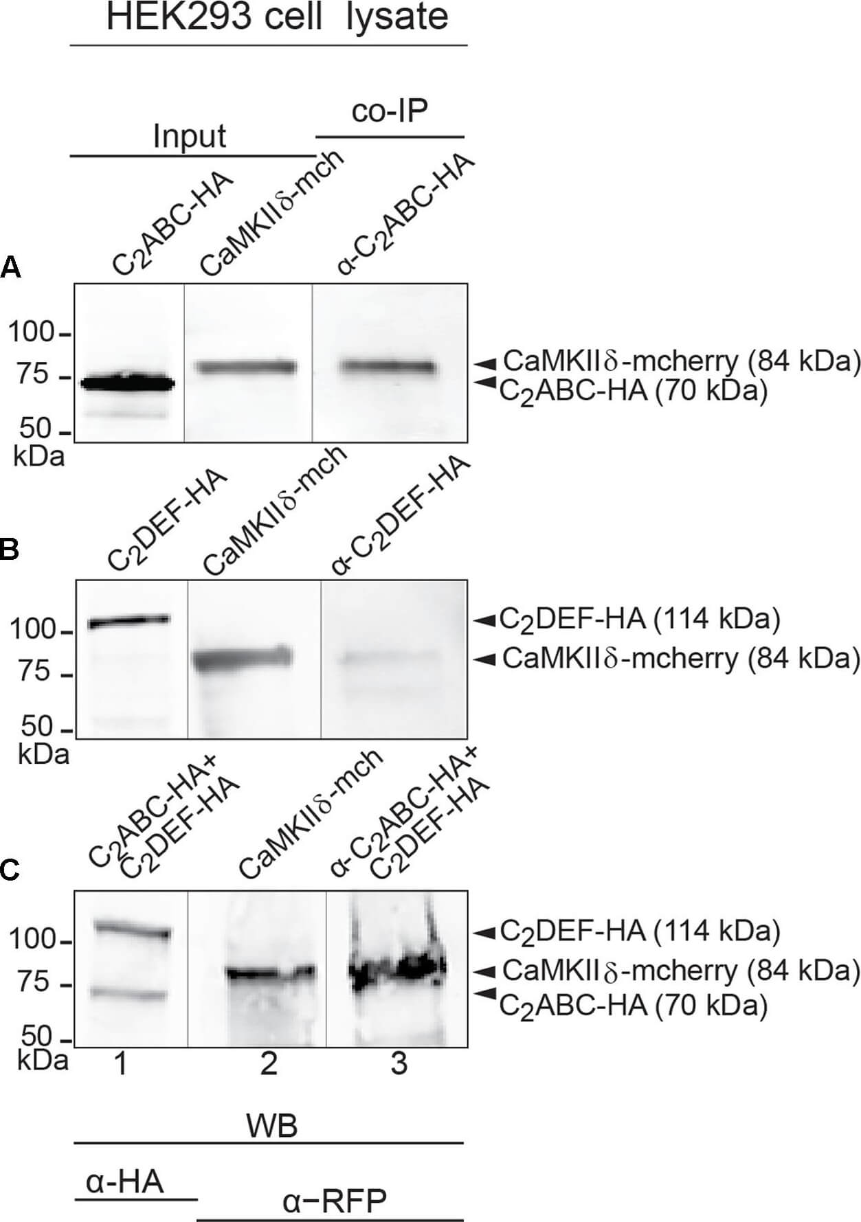
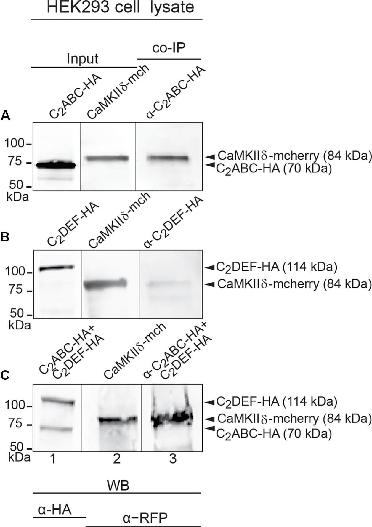
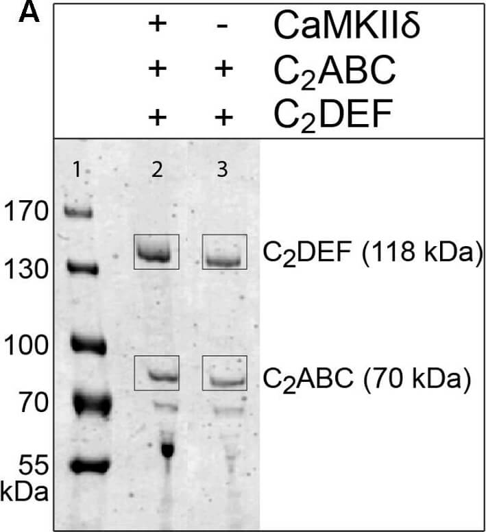
Immunoprecipitation and western blot show interaction of otoferlin with CaMKIIdelta. (A-C) Two HA-tagged mouse otoferlin fragments, C2ABC (aa 1-632 in NP_001093865, 70 kDa) and C2DEF (aa 933-1920, 114 kDa) were co-transfected with mcherry-tagged mouse CaMKIIdelta into HEK293 cells. Transfections were performed either with otoferlin C2ABC and CaMKIIdelta (A, Input Lane 1 and 2), otoferlin C2DEF and CaMKIIdelta (B, Input Lan
Immunoprecipitation and western blot show interaction of otoferlin with CaMKIIdelta. (A-C) Two HA-tagged mouse otoferlin fragments, C2ABC (aa 1-632 in NP_001093865, 70 kDa) and C2DEF (aa 933-1920, 114 kDa) were co-transfected with mcherry-tagged mouse CaMKIIdelta into HEK293 cells. Transfections were performed either with otoferlin C2ABC and CaMKIIdelta (A, Input Lane 1 and 2), otoferlin C2DEF and CaMKIIdelta (B, Input Lane 1 and 2) or in the presence of both C2ABC and C2DEF fragments and CaMKIIdelta (C, Input Lane 1 and 2). Co-immunoprecipitations of C2ABC-HA and C2DEF-HA were conducted from HEK293 cell lysates using anti-HA antibodies. CaMKIIdelta-mcherry was detected in the eluate using an anti-RFP (red fluorescent protein) antibody (A-C, Lane 3), indicating that CaMKIIdelta co-precipitated with recombinant otoferlin fragments. Figure provided by CiteAb. Source: Front Synaptic Neurosci, PMID: 29046633.
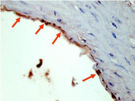
Rocklands Affinity Purified anti-HA epitope tag polyclonal antibody detects HA tagged recombinant proteins by IHC on formalin fixed paraffin embedded tissue. Arrowheads point to expression of HA tagged proteins in endothelial cells of mouse aorta. Sections of 4 µm were prepared from representative paraffin blocks. Sections were then deparaffinized and rehydrated with xylene and alcohol. Citrate buffer antigen retrieval was performed for 30 min in a boiling jar. Anti-HA was diluted in blocking buffer at 1:2,000 and reacted at 4 C overnight followed by signal detection using horseradish peroxidase with DAB as the chromogenic substrate. Tissue was counterstained with Mayers hematoxylin. Personal Communication, Behzad Yeganeh,U. Manitoba, Winnipeg, Canada.
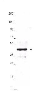
Anti-HA epitope tag polyclonal antibody detects HA-tagged recombinant proteins by western blot. Polyclonal Rabbit anti-HA epitope tag, at a 1:2,000 dilution, was used to detect 1.0 µg of 12-Epitope Tag Protein Marker Lysate (p/n MB-301-0100) containing the HA epitope tag. A 4-20% gradient gel was used to resolve the protein by SDS-PAGE. The lysate was transferred to nitrocellulose using standard methods. After blocking, the membrane was probed with Rocklands anti-HA tag antibody for 1 h at room temperature followed by washes and reaction with a 1:20,000 dilution of IRDye 800 conjugated Gt-a-Rabbit IgG (H&L) MX10 (code 611-132-122) for 30 min at room temperature. LICORs Odyssey Infrared Imaging System was used to scan and process the image. Other detection systems will yield similar results.
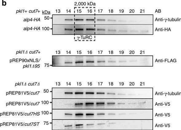
Kinesin-5 Cut7 binds the gamma-TuRC MTOC.(a) Kinesin-5 and kinesin-14 constructs used in Fast Protein Liquid Chromatography. V5-tagged Cut7 and two truncation constructs were used, in addition to one FLAG-Pkl1 truncated construct that retains full Pkl1 activity. Cut7 constructs are V5-tagged full-length Cut7 (aa 1-1,085), Cut7-Head-Stalk (Cut7HS, aa 1-888) and Cut7-Stalk-Tail (Cut7-ST, aa 443-1,085). (b) Western blot profiles of whole-cell extracts fractionated by Separose 6 using FPLC. (c) Western blots of Cut7 constructs immunoprecipitated from whole-cell extracts using anti-V5 magnetic beads with empty strain negative controls. (d) Cartoon diagram of 6-His tagged Pkl1 Tail peptide co-immunoprecipitation assay using magnetic beads with His affinity and FPLC fraction 15. (e) Pkl1 Tail peptide co-immunoprecipitation of gamma-TuRC core subunits and V5-Cut7ST using a short Pkl1 Tail peptide (PgammaT). Mutated peptide PgammaM has significantly reduced interaction with the fission yeast gamma-TuRC. The anti-HA antibody detects the HA-tagged gamma-TuRC protein Alp4. Figure provided by CiteAb. Source: Nat Commun, PMID: 25348260.
* Mehrwertsteuer und Versandkosten nicht enthalten. Irrtümer und Preisänderungen vorbehalten