Human Myosin Light Chain phospho peptide corresponding to a region near the amino terminus of the human smooth/non-muscle form of myosin regulatory light chain conjugated to Keyhole Limpet Hemocyanin (KLH).
This phospho specific polyclonal antibody was tested by ELISA, immunohistochemistry, and immunoblotting. Immunoblotting was used to show reactivity with unstimulated and stimulated cardiac myocytes. The antibody was also reactive with the phosphorylated
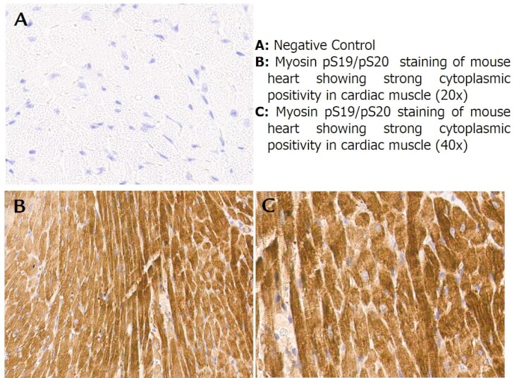
Immunohistochemistry with anti-myosin pS19/pS20 antibody showing strong cytoplasmic staining of myocytes in mouse heart muscle 20x and 40x (B & C). Staining was performed on Leica Bond system using the standard protocol. Formalin fixed/paraffin embedded tissue sections were subjected to antigen retrieval and then incubated with rabbit anti-myosin pS19/pS20 antibody at 1:100 dilution for 60 minutes. Biotinylated Anti-rabbit secondary antibody was used to detect primary antibody. The reaction was developed using streptavidin-HRP conjugated compact polymer system and visualized with chromogen substrate, 33-diamino-benzidine substrate (DAB). The sections were then counterstained with hematoxylin to detect cell nuclei.
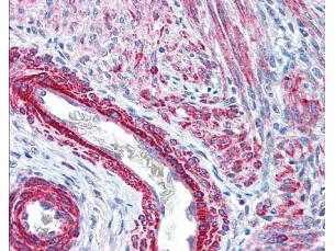
Rocklands affinity purified anti-Monophosphorylated RLC Smooth and Non-Muscle Myosin pS19/20 antibody was used at 2.5 µg/ml to detect signal in a variety of tissues including multi-human, multi-brain and multi-cancer slides. This image shows strong staining of both vascular and myometrial smooth muscle cells of the uterus. Tissue was formalin-fixed and paraffin embedded. The image shows localization of the antibody as the precipitated red signal, with a hematoxylin purple nuclear counterstain. Personal Communication, Tina Roush, LifeSpanBiosciences, Seattle, WA.
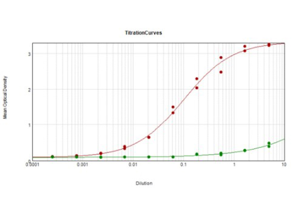
ELISA Results of Rabbit Anti-Myosin pS19/pS20 Antibody tested against BSA-conjugated peptide of immunizing peptide. Each well was coated in duplicate with 0.1µg of Myosin pS19/pS20 [Red Line] and Myosin S19/S20 [Green Line]. The starting dilution of antibody was 5µg/ml and the X-axis represents the Log10 of a 3-fold dilution. This titration is a 4-parameter curve fit where the IC50 is defined as the titer of the antibody. Assay performed using Goat anti-Rabbit IgG Antibody Peroxidase Conjugated (Min X Bv Ch Gt GP Ham Hs Hu Ms Rt & Sh Serum Proteins) (p/n 6
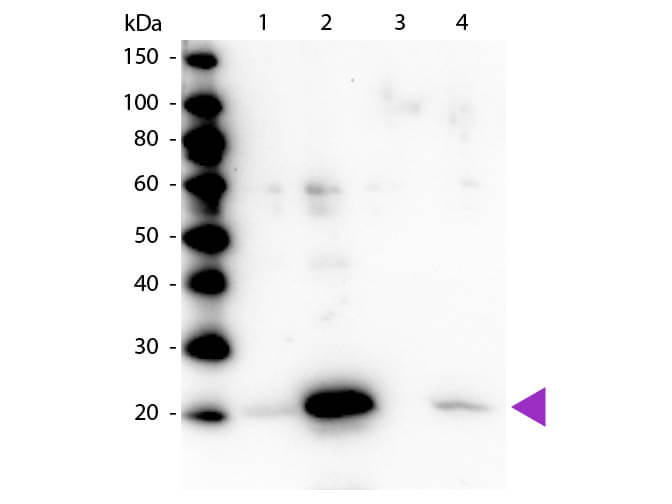
Western blot of Rabbit Anti-Myosin pS19/pS20 primary antibody. Lane 1: Regulatory Light Chain Non-Phospho recombinant protein. Lane 2: Regulatory Light Chain Phospho recombinant protein. Lane 3: Smooth Muscle Non-Phospho recombinant protein. Lane 4: Smooth Muscle Phospho recombinant protein. Load: 50 ng per lane. Primary antibody: Myosin pS19/pS20 primary antibody at 1:1,000 overnight at 4°C. Secondary antibody: Peroxidase rabbit secondary antibody at 1:40,000 for 60 min at RT. Blocking: MB-070 for 30 min at RT. Predicted/Observed size: 20 kDa, 20 kDa for Regulatory Light Chain Phospho. Other band(s): None.
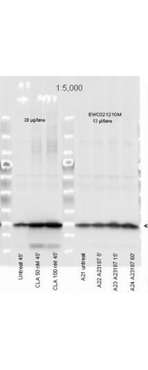
Affinity Purified Phospho specific antibody to Monophosphorylated Regulatory Light Chain of Smooth and Non-muscle Myosin at pS19/pS20 was used at a 1:5000 dilution to detect myosin light chain by Western blot. Either 13µL or 20 µg of a mouse cardiac myocyte lysate was loaded on a 4-20% Criterion gel for SDS-PAGE. Samples were either mock-treated or CLA-treated, as indicated. After washing, a 1:5,000 dilution of HRP conjugated Gt-a-Rabbit IgG (611-103-122) preceded color development using Amershams substrate system. Other detection methods will yield similar results. Data courtesy of the Alliance for Cellular Signaling (http://www.signaling-gateway.org).
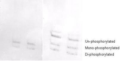
Affinity purified phosphospecific antibody to phosphorylated regulatory light chain of smooth and non-muscle Myosin at pS19/pS20 was used at a 1:1000 dilution to detect myosin light chain by Western blot on 3T3 cell lysates. A standard urea/glycerol gel without SDS was used to separate phospho forms of regulatory light chain according to mass to charge ratios. In Panel A on the left, reactivity of Rocklands phosphospecific antibody is shown. In Panel B on the right, reactivity of commercially available pan reactive antibody that detects both un-phosphorylated and phosphorylated forms of regulatory light chain is shown. Rocklands phosphospecific antibody detects both mono-phosphorylated (pSer20 Mono-P-RLC) and di-phosphorylated (pThr19-pSer20 Di-P-RLC) regulatory light chain. Personal communication. J. Stull. UT Southwestern Medical Center.
* Mehrwertsteuer und Versandkosten nicht enthalten. Irrtümer und Preisänderungen vorbehalten