This affinity purified antibody was prepared from whole rabbit serum produced by repeated immunizations with a synthetic peptide corresponding to an internal region near aa 575-600 of mouse Hif3 a (Hypoxia Inducible Factor).
Konjugation:
Unconjugated
Alternative Synonym:
rabbit anti-Hif3a antibody, Hif3 alpha antibody, Hif3alpha antibody, Hypoxia Inducible Factor 3-alpha antibody, Hypoxia inducible factor 3 alpha subunit antibody, Inhibitory PAS domain protein antibody, IPAS antibody, HIF-3-alpha, Basic-helix-loop-helix-PAS protein MOP7, HIF3-alpha-1, Neonatal and embryonic PAS protein
0.02 M Potassium Phosphate, 0.15 M Sodium Chloride, pH 7.2
Formulierung:
Liquid (sterile filtered)
Target-Kategorie:
Mouse
Antibody Type:
Primary Antibody
Application Verdünnung:
ELISA: 1:14,000 - 1:80,000, WB: 1:1,000 - 1:8,000
Anwendungsbeschreibung:
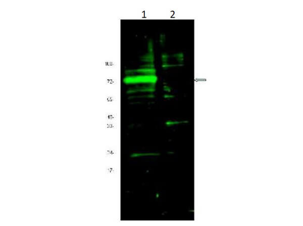
This affinity-purified antibody has been tested for use in ELISA and western blot. Specific conditions for reactivity should be optimized by the end user. Expect a band approximately 72 kDa in size corresponding to Hif3 alpha protein by western blotting
Western blot using Rocklands Affinity Purified Anti-Hif3A antibody shows detection of a band ~72 kDa corresponding to mouse Hif3A [arrowhead]. Approximately 10 µg of a CoCl2 treated 3T3 cell lysate [lane 1] and control 3T3 cell lysate (p/n W10-000-358) [lane 2] were separated by 4-20% SDS-PAGE and transferred onto nitrocellulose. Treatment of exponentially growing 3T3 cells with 130 µM CoCl2 for 6 h at 37 C effectively mimics hypoxia. After blocking the membrane was probed overnight at 4 C with the primary antibody diluted to 1:1,600. The membrane was washed and reacted with a 1:10,000 dilution of IRDye(TM)800 conjugated Gt-a-Rabbit IgG [H&L] MX (p/n 611-132-122) for 45 min at room temperature. IRDye(TM)800 fluorescence image was captured using the Odyssey Infrared Imaging System developed by LI-COR. IRDye is a trademark of LI-COR, Inc. Other detection systems will yield similar results.
* Mehrwertsteuer und Versandkosten nicht enthalten. Irrtümer und Preisänderungen vorbehalten