This affinity purified antibody was prepared from whole rabbit serum produced by repeated immunizations with a synthetic peptide corresponding to an internal region near amino acids 290-320 of human IRS1 protein.
This affinity purified antibody has been tested for use in ELISA and western blot. Specific conditions for reactivity should be optimized by the end user. Expect a band approximately 130 kDa in size corresponding to phosphorylated IRS1 protein by western
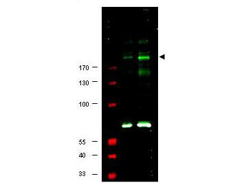
Western blot using Rocklands Affinity Purified anti-IRS1 pS307 antibody shows detection of a band at ~180 kDa believed to represent phosphorylated IRS1 (arrowhead). Lane 1 shows staining of human 293 cell lysate (p/n W09-000-365). Lane 2 shows staining of 293 cell lysate prepared from cells serum-starved for 18 h followed by treatment with 5 µg/ml of anisomysin for 30 min. The pronounced staining of the band at 180 kDa is not seen when the antibody was pre-incubated with immunizing peptide prior to reaction (data not shown). The identity of the intensely reactive bands at ~70 kD in both lane 1 and 2 is unknown, although these bands were also competed out by pre-incubation with the immunizing peptide. Approximately 25 µg of each lysate was separated on a 4-20% Tris-Glycine gel by SDS-PAGE and transferred onto nitrocellulose. After blocking with 5% goat serum, 0.5% BLOTTO in PBS, the membrane was probed with the primary antibody diluted to 1:250. Reaction occurred overnight at 4 C followed by washes and reaction with a 1:10,000 dilution of IRDye(TM)800 conjugated Gt-a-Rabbit IgG [H&L] MX (611-132-122) for 45 min at room temperature (800 nm channel, green). Molecular weight estimation was made by comparison to prestained MW markers in lane M (700 nm channel, red). IRDye(TM)800 fluorescence image was captured using the Odyssey Infrared Imaging System developed by LI-COR. IRDye is a trademark of LI-COR, Inc. Other detection systems will yield similar results.
* Mehrwertsteuer und Versandkosten nicht enthalten. Irrtümer und Preisänderungen vorbehalten