This affinity purified antibody was prepared from whole rabbit serum produced by repeated immunizations with a synthetic peptide corresponding to an internal region of the WHIP1 protein. The immunogen sequence shows 100% homology to human WHIP1 (isoform 1) and WHIP2 (isoform 2) with predicted molecular weights of 72.2 kDa and 69.5 kDa, respectively. The immunogen sequence also shows 100% homology to WHIP1 from mouse, rat and monkey sequences. Reactivity with WHIP proteins from other sources is not known, but is likely due to reported homologies.
Konjugation:
Unconjugated
Alternative Synonym:
rabbit anti-WHIP antibody, ATPase WRNIP 1 antibody, ATPase WRNIP1 antibody, Werner helicasae interacting protein 1 antibody, WRNIP-1
This affinity purified antibody has been tested by WB and ELISA. Anti-WHIP is useful in western blotting against HEK293 whole cell lysates. Dilutions for western blotting represent a starting point dilution and further optimization may be required. The a
Western blot analysis is shown using Rocklands anti-Human WHIP antibody with and without pre-incubation with blocking peptide. Testing was performed on antiserum prior to affinity purification. Peptide competition (left) blocks the specific staining, whereas the control (right) shows staining of a strong dominant band corresponding to human WHIP1. ~30µg of HEK293 lysate was loaded per lane for 4-20% gradient SDS-PAGE. Comparison to a molecular weight marker (not shown) indicates a band of ~96.0 kDa is detected. The blot was incubated with a 1:1000 dilution of the antibody at room temperature for 2 h followed by detection using IRDye 800 labeled Goat-a-Rabbit IgG [H&L] MX10 (611-132-122) diluted 1:5,000 for 45 min. IRDye 800 fluorescence image was captured using the Odyssey Infrared Imaging System developed by LI-COR. IRDye is a trademark of LI-COR, Inc. Other systems will yield similar results.
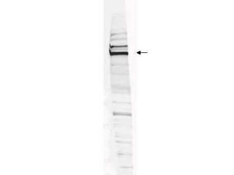
Western blot analysis is shown using Rocklands Affinity Purified anti-Human WHIP antibody to detect Human WHIP present in a HEK293 whole cell lysate. ~30µg of lysate was loaded per lane for 4-20% gradient SDS-PAGE. Comparison to a molecular weight marker (not shown) indicates a primary band of ~96.0 kDa is detected. The identity of the minor band migrating at a slightly higher molecular weight is unknown, but may represent an alternate isoform of WHIP or post translational modification of the WHIP protein. See Figure 2 for the results of peptide competition experiments. The blot was incubated with a 1:200 dilution of the antibody at room temperature for 2 h followed by detection using IRDye 800 labeled Goat-a-Rabbit IgG [H&L] MX10 (611-132-122) diluted 1:5,000 for 45 min. IRDye 800 fluorescence image was captured using the Odyssey Infrared Imaging System developed by LI-COR. IRDye is a trademark of LI-COR, Inc. Other detection systems will yield similar results.
* Mehrwertsteuer und Versandkosten nicht enthalten. Irrtümer und Preisänderungen vorbehalten