This affinity purified antibody was prepared from whole rabbit serum produced by repeated immunizations with a synthetic peptide corresponding to an internal region near amino acids 350-375 of Human E2F-1.
This affinity purified antibody has been tested for use in ELISA, immunohistochemistry and by western blot. Specific conditions for reactivity should be optimized by the end user. Expect a band approximately 47 kDa in size corresponding to phosphorylated
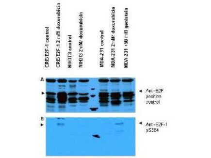
Western blot using Rocklands Affinity Purified anti-E2F-1 pS364 antibody shows detection of a band at ~47 kDa corresponding to phosphorylated E2F-1 in induced cell lysates. Panel A shows reactivity using a control antibody reactive to all forms of E2F (arrowheads). Panel B shows specific reactivity against phosphorylated E2F-1 (arrowheads) using our anti-E2F-1 pS364 antibody. Lysates are as follows: CRE/E2F-1 are CRE cells derived from mouse NIH3T3 line transfected with human E2F-1, NIH-3T3 used as a negative control, and MDA-MB-231 cells are a human breast cancer line. As indicated each lysate was prepared from untreated cells and cells treated with 2 µM Doxorubicin used as a DNA damaging agent. In addition the MDA-MB-231 cells were also treated with genistein, a mild DNA damaging agent. The figure shows the same membrane first probed with the anti-E2F-1 pS364 antibody used at a 1:250 dilution, then stripped and re-probed with the pan E2F antibody used as a positive control. The positive control antibody clearly shows an E2F-1 band in all human cell lines, but not mouse cells. Treatment with doxorubicin increases the expression of E2F-1 as shown in Panel A. After film development, images were overlapped to confirm that specific anti-E2F-1 pS364 staining shown treated human cells in Panel B specifically aligns with E2F-1 staining shown in Panel A. Blots can be processed with HRP conjugated Gt-a-Rabbit IgG MX10 611-103-122 for 45 min at room temperature for ECL detection. Personal Communication, XiaoHe Yang, Univ. Oklahoma.
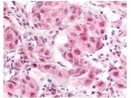
Rocklands Affinity Purified anti- E2F-1 pS364 antibody was used at a 10 µg/ml to detect nuclear and occasionally cytoplasmic signal in a variety of tissues including multi-human, multi-brain and multi-cancer slides. Within the multi-tumor block, the antibody showed variable levels of nuclear staining between individual tumors, with some tumors showing strong staining. This image shows E2F-1 pS364 staining of human breast carcinoma. Tissue was formalin-fixed and paraffin embedded. Personal Communication, Tina Roush, LifeSpanBiosciences, Seattle, WA.
* Mehrwertsteuer und Versandkosten nicht enthalten. Irrtümer und Preisänderungen vorbehalten