This affinity-purified antibody was prepared from whole rabbit serum produced by repeated immunizations with a synthetic peptide corresponding to an C-Terminal region near amino acids 1625-1650 of Human ROBO-1.
Konjugation:
Unconjugated
Alternative Synonym:
rabbit anti-ROBO1 antibody, ROBO 1, ROBO-1, hROBO-1, Roundabout homolog 1, Deleted in U twenty twenty, DUTT1, DUTT-1
0.02 M Potassium Phosphate, 0.15 M Sodium Chloride, pH 7.2
Formulierung:
Liquid (sterile filtered)
Target-Kategorie:
Human
Antibody Type:
Primary Antibody
Application Verdünnung:
ELISA: 1:30,000 - 1:160,000, IHC: 2 µg/ml to 10 µg/ml, IF Microscopy: User Optimized, WB: 1:500 - 1:3,000
Anwendungsbeschreibung:
This affinity purified antibody has been tested for use in ELISA, western blot, and immunohistochemistry. It may be suitable for immunofluorescence and IP. Specific conditions for reactivity should be optimized by the end user. Expect a band at ~181 kDa
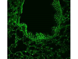
1/50 staining mouse lung tissue sections (adult, frozen 100µm wholemount sections) by IHC-Fr. The tissue was paraformaldehyde fixed and permeabilized with triton x-100 before incubation with the antibody for 16 hours at 4C.
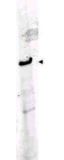
Western blot using Rocklands Affinity Purified anti-ROBO-1 antibody shows detection of a band at ~181 kDa corresponding to ROBO-1 present in mouse brain lysate (p/n W10-000-T004) (arrowhead). Approximately 35 µg of lysate was separated by 4-8% SDS-PAGE and transferred onto nitrocellulose. After blocking the membrane was probed with the primary antibody diluted to 1:1,000. Reaction occurred 2h at room temperature followed by washes and reaction with a 1:10,000 dilution of IRDye(TM)800 conjugated Gt-a-Rabbit IgG [H&L] MX (p/n 611-132-122) for 45 min at room temperature. IRDye(TM)800 fluorescence image was captured using the Odyssey Infrared Imaging System developed by LI-COR. IRDye is a trademark of LI-COR, Inc. Other detection systems will yield similar results.
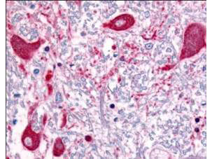
Rocklands Affinity Purified anti-ROBO1 antibody was used at a concentration of 5 µg/ml to detect ROBO1 in a variety of tissues including multi-human, multi-brain and multi-cancer slides. This image shows staining of human brain tissue. Tissue was formalin-fixed and paraffin embedded. Personal Communication, Tina Roush, LifeSpanBiosciences, Seattle, WA.
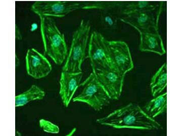
Staining of ROBO1 in undifferentiated, immortalized human podocytes by Immunocytochemistry/ Immunofluorescence. Cells were fixed with 2% paraformaldehyde and 4% sucrose at room temperature for 10 minutes. The cells were then washed once with PBS, permeabilized with 0.3% Triton X-100 for 10 minutes and incubated with blocking solution (2% FCS, 2% BSA, 0.2% fish gelatin) for 30 minutes, before further incubation with primary Ab for 1 hour. An Alexa Fluor 488 goat anti-rabbit IgG secondary antibody was used at a dilution of 1/200. DAPI was used for nuclear counterstaining. Image from Lindenmeyer MT et al. Systematic Analysis of a Novel Human Renal Glomerulus-Enriched Gene Expression Dataset. PLoS One. 2010 July 12,5(7):e11545, Fig 5.
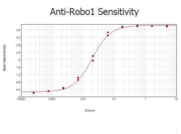
ELISA results of purified Rabbit anti-Robo-1 Antibody tested against BSA-conjugated peptide of immunizing peptide. Each well was coated in duplicate with 0.1µg of conjugate. The starting dilution of antibody was 5µg/ml and the X-axis represents the Log10 of a 3-fold dilution. This titration is a 4-parameter curve fit where the IC50 is defined as the titer of the antibody. Assay performed using 3% fish gel, Goat anti-Rabbit IgG Antibody Peroxidase Conjugated (Min X Bv Ch Gt GP Ham Hs Hu Ms Rt & Sh Serum Proteins) (p/n 611-103-122) and TMB ELISA Peroxidase Substrate (p/n TMBE-1000).
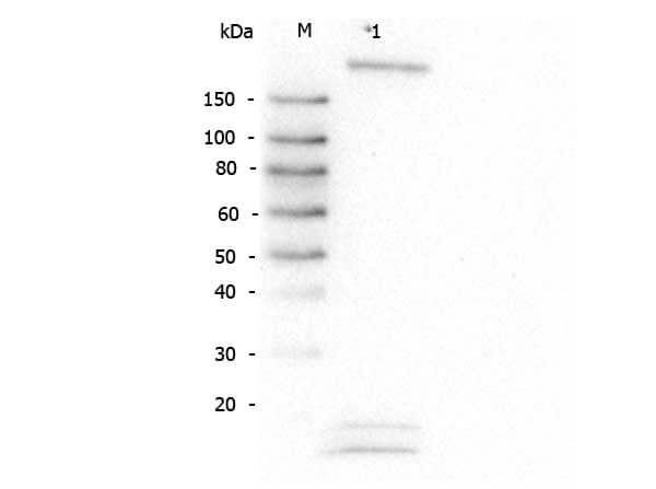
Western Blot of Rabbit anti-Robo-1 antibody. Lane M: Super Signal Molecular Weight Marker. Lane 1: HeLa WCL (p/n W09-000-364). Load: 30 µg lysate. Primary antibody: Robo-1 antibody at 1:1,000 for overnight at 4C. Secondary antibody: Peroxidase rabbit secondary antibody (p/n 611-103-122) at 1:40,000 for 30 min at RT. Block: Blocking Buffer for Fluorescent Western Blotting (p/n MB-070) for 30 min at RT. Predicted
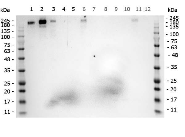
Western Blot of Rabbit anti-ROBO1 antibody. Marker: Opal Pre-stained ladder (p/n MB-210-0500). Lane 1: HEK293 lysate (p/n W09-000-365). Lane 2: HeLa Lysate (p/n W09-000-364). Lane 3: MCF-7 Lysate (p/n W09-000-360). Lane 4: Jurkat Lysate (p/n W09-000-370). Lane 5: A431 Lysate (p/n W09-000-361). Lane 6: A549 Lysate (p/n W09-001-372). Lane 7: LNCap Lysate (p/n W09-001-GJ9). Lane 8: MOLT-4 Lysate (p/n W09-001-GK2). Lane 9: Ramos Lysate (p/n W09-000-GK4). Lane 10: Raji Lysate (p/n W09-001-368). Lane 11: A-172 Lysate (p/n W09-001-GL5). Lane 12: NIH/3T3 Lysate (p/n W10-000-358). Load: 35 µg per lane. Primary antibody: ROBO1 antibody at 1ug/mL overnight at 4C. Secondary antibody: Peroxidase rabbit secondary antibody (p/n 611-103-122) at 1:30,000 for 60 min at RT. Blocking Buffer: 1% Casein-TTBS (p/n MB-082) for 30 min at RT. Predicted/Observed size: 181kDa for ROBO1.
* Mehrwertsteuer und Versandkosten nicht enthalten. Irrtümer und Preisänderungen vorbehalten