This affinity purified antibody was produced from monospecific rabbit serum by repeated immunizations with a synthetic peptide corresponding to an internal region near amino acids 30-60 of human Gli-3 protein.
Konjugation:
Unconjugated
Alternative Synonym:
Rabbit anti-GLI-3 antibody, Transcriptional activator GLI3, Gli 3, GLI3 form of 190 kDa, GLI3 form of 83 kDa
0.02 M Potassium Phosphate, 0.15 M Sodium Chloride, pH 7.2
Formulierung:
Liquid (sterile filtered)
Target-Kategorie:
Human
Antibody Type:
Primary Antibody
Application Verdünnung:
ELISA: 1:6,000 - 1:30,000, IHC: 0.5 mg/ml - 5 µg/ml, IF Microscopy: User Optimized, WB: 1:500 - 1:2,000
Anwendungsbeschreibung:
This antibody has been tested for use in ELISA, immunohistochemistry, immunofluorescence, and western blot. Specific conditions for reactivity should be optimized by the end user. Detection of Gli-3 by western blot may be enhanced if nuclear extracts are
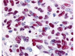
Immunohistochemistry of Rabbit anti-Gli-3 antibody. This image tissue: human glioblastoma. Specific staining was also noted in tissue from adrenal, brain, glioblastoma, colon, heart, kidney, lung, liver, skeletal muscle, ovary, pancreas, placenta, skin, spleen, stomach, testes, thymus, thyroid, tonsil and uterus. Fixation: formalin fixed paraffin embedded. Antigen retrieval: not required. Primary antibody: Gli-3 antibody at 0.625 µg/ml for 1 h at RT. Secondary antibody: Peroxidase rabbit secondary antibody at 1:10,000 for 45 min at RT. Localization: Gli-3 is nuclear and smooth muscle. Staining: Gli-3 as precipitated red signal with hematoxylin purple nuclear counterstain.
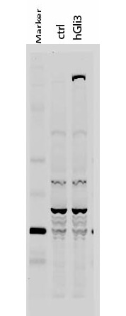
Western Blot of Rabbit anti-Gli-3 antibody. Lane 1: 50 kDa molecular weight marker. Lane 2: 293T cells transfected with CrkL-Flag. Lane 3: 293T cells transfected with human Gli-3. Load: 35 µg per lane. Primary antibody: Gli-3 antibody at 1:400 for overnight at 4C. Secondary antibody: IRDye800(TM) rabbit secondary antibody at 1:10,000 for 45 min at RT. Block: 5% BLOTTO overnight at 4C. Predicted/Observed size: 170-190 kDa for hGli-3. Other band(s): Non specific background ~60kDa.
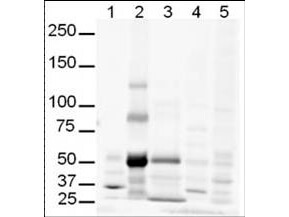
Western Blot of Rabbit anti-Gli-3 antibody. Lane 1: human brain whole cell lysate. Lane 2: human lung whole cell lysate. Lane 3: human spleen whole cell lysate. Lane 4: mouse brain whole cell lysate (p/n W10-000-T004). Lane 5: mouse lung whole cell lysate (p/n W10-000-MQ1). Load: 20 µg per lane. Primary antibody: Gli-3 antibody at 1:500 for overnight at 4C. Secondary antibody: IRDye800(TM) rabbit secondary antibody at 1:10,000 for 45 min at RT. Block: 5% BLOTTO overnight at 4C. Predicted/Observed size: Isoforms at ~170-190kDa and ~80kDa. Lane 2 shows what may be truncated Gli-3 (~80kDa). Other band(s): The strong band at ~50 kDa is unknown.
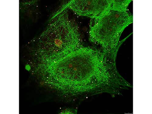
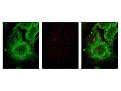
Immunofluorescence Microscopy of Rabbit anti-Gli-3 antibody. Tissue: MCF-7 cell. Antigen retrieval: not required. Primary antibody: Gli-3 antibody and Anti-alpha-Tubulin at 5 µg/mL for 1 h at RT. Secondary antibody: Fluorescein secondary antibody at 1:10,000 for 45 min at RT. Localization: Gli-3 is nuclear. Staining: Gli-3 staining as red fluorescent signal and Anti-alpha-Tubulin staining as green fluorescent signal using STED.
Immunofluorescence Microscopy of Rabbit anti-Gli-3 antibody. Tissue: MCF-7 cell. Antigen retrieval: not required. Primary antibody: Gli-3 antibody and Anti-alpha-Tubulin at 5 µg/mL for 1 h at RT. Secondary antibody: Fluorescein secondary antibody at 1:10,000 for 45 min at RT. Localization: Gli-3 is nuclear. Staining: Image (1) shows alpha-Tubulin staining as green fluorescent signal. Image (2) shows Gli-3 staining as red fluorescent signal and Images (3) shows both antibodies fluorescing using STED microscopy.
* Mehrwertsteuer und Versandkosten nicht enthalten. Irrtümer und Preisänderungen vorbehalten