Anti-Tubulin Loading Control Antibody was prepared from whole rabbit serum produced by repeated immunizations with a synthetic peptide corresponding to the C-Terminal region near amino acids 425-451 of Human alpha Tubulin.
Anti-Tubulin Antibody has been tested for use in ELISA, immunofluorescence, and western blot. Specific conditions for reactivity should be optimized by the end user. Expect a band at ~50 kDa in size corresponding to alpha tubulin by western blotting in m
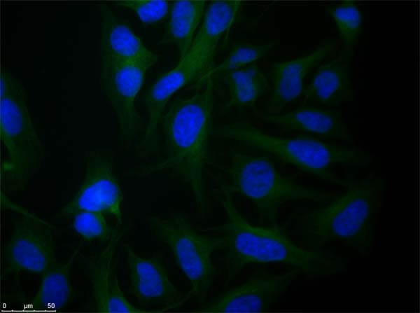
Immunofluorescence microscopy of Rabbit Anti-alpha-Tubulin antibody using HeLa cells fixed with PFA. Anti-alpha-Tubulin Antibody was used at 1 µg/mL, O/N at 4°C. Secondary antibody: Anti-RABBIT IgG DyLight(TM) 488 Conjugated Preadsorbed (p/n 611-741-127) at 2 ug/ml for 1 h at RT. Localization: TUBA1B is the major constituent of microtubules in the cytoplasm. Staining: Tubulin as green fluorescent signal with DAPI (blue) nuclear counterstain.
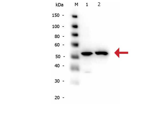
Western Blot of Rabbit anti-alpha-Tubulin antibody. Lane 1: HeLa WCL (p/n W09-000-364). Lane 2: NIH/3T3 WCL (p/n W10-000-358). Load: 10 µg per lane. Primary antibody: alpha-Tubulin antibody at 1:1,000 for overnight at 4C. Secondary antibody: Peroxidase rabbit secondary antibody (p/n 611-103-122) at 1:40,000 for 30 min at RT. Block: Blocking Buffer for Fluorescent Western Blotting (p/n MB-070) for 30 min at RT. Predicted/Observed size: 50 kDa, 50 kDa for alpha-Tubulin. Other band(s): N/A.
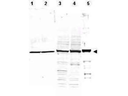
Western Blot of Rabbit Anti-Alpha Tubulin Antibody. Lane 1: whole cell lysates from mouse brain (p/n W10-000-T004). Lane 2: rat brain (p/n W12-000-T077). Lane 3: A431 cells (p/n W09-000-361). Lane 4: Jurkat cells (p/n W09-001-370). Lane 5: HeLa cells (p/n W09-000-364). Load: 35 µg per lane. Primary antibody: Alpha Tubulin antibody at 1:1,200 for overnight at 4C. Secondary antibody: IRDye800(TM) rabbit secondary antibody at 1:10,000 for 45 min at RT. Block: 5% BLOTTO (p/n B501-0500) overnight at 4C. Predicted/Observed size: ~50 kDa corresponding to alpha tubulin (arrowhead). Other band(s): none.
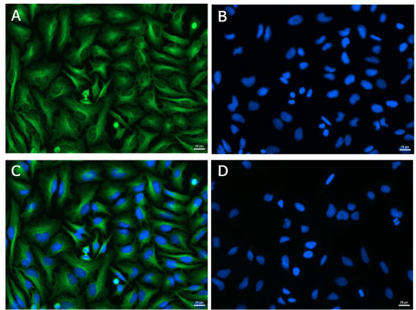
Immunofluorescence of Rabbit Anti-Alpha Tubulin Antibody. Cell line: HeLa. Primary Antibody: Alpha Tubulin (p/n 600-401-880) at 4.4 µg/mL (1:250) for 1hr at RT. Secondary Antibody: Goat Anti-Rabbit DyLight(TM) 488 (p/n 611-141-121) at 1 µg/mL (1:1000) overnight at 4 C. Fixative: Ice Cold Methanol. Permeabilization: Ice Cold Methanol. Nuclear stain: Hoechst 33342. Expected Localization: Cytoplasmic. Image: A) Alpha Tubulin, B) Nuclear Stain, C) Merge, D) Secondary Only Control.
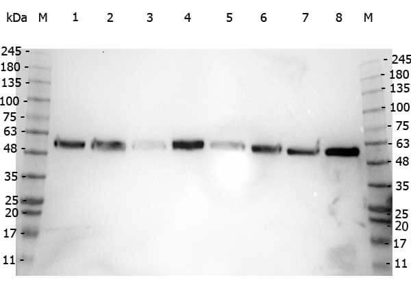
Western Blot of Rabbit anti-Alpha-Tubulin antibody. Marker: Opal Pre-stained ladder (p/n MB-210-0500). Lane 1: HEK293 lysate (p/n W09-000-365). Lane 2: HeLa Lysate (p/n W09-000-364). Lane 3: MCF-7 Lysate (p/n W09-000-360). Lane 4: Jurkat Lysate (p/n W09-000-370). Lane 5: A431 Lysate (p/n W09-000-361). Lane 6: LNCaP Lysate (p/n W09-001-GJ9). Lane 7: A-172 Lysate (p/n W09-001-GL5). Lane 8: NIH/3T3 Lysate
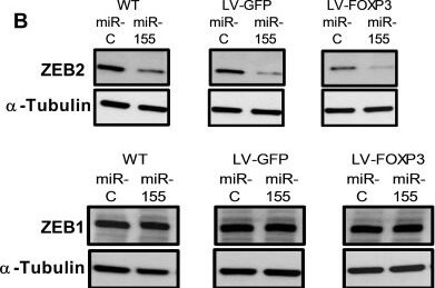
miR-155 and FOXP3 down regulate endogenous ZEB2 in human breast cancer cells resulting in altered levels of EMT markers Vimentin and E-cadherin(A) Relative abundance of ZEB2 and ZEB1 protein in WT, GFP or FOXP3 overexpressing BT549 cells transfected with miR-155 or miR-control. Relative abundance of protein was determined by quantitation of the abundance of ZEB2 or ZEB1 proteins normalised to reference protein alpha-Tubulin by western blot analysis. Quantitation of bands was carried out using Image J software. Mean + SD plotted. Students t test ***P < 0.001. ZEB1 protein expression as above. n = 3 experiments. (B) ZEB2 and ZEB1 protein in WT, GFP or FOXP3 overexpressing BT549 cells transfected with miR-155 or miR-control by western blot. Representative western blot shown. (C) Relative abundance of Vimentin and E-cadherin protein in WT, GFP or FOXP3 overexpressing BT549 cells transfected with miR-155 or miR-control. Relative abundance of protein was determined by quantitating the abundance of E-cadherin or Vimentin proteins and normalising to reference protein beta-Actin by western blot analysis. Quantitation of bands was carried out using Image J software. Mean + SD plotted. Students t test ***P < 0.001, **P < 0.01. n = 3 experiments. (D) Vimentin and E-cadherin protein in WT, GFP or FOXP3 overexpressing BT549 cells transfected with miR-155 or miR-control analysed by western blot. Representative western blot shown. Figure provided by CiteAb. Source: Oncotarget, PMID: 29963231.
* Mehrwertsteuer und Versandkosten nicht enthalten. Irrtümer und Preisänderungen vorbehalten