This affinity purified antibody was prepared from whole rabbit serum produced by repeated immunizations with a synthetic peptide corresponding to an internal region near aa 305-330 of S. cerevisiae CHK1.
This affinity purified antibody has been tested for use in ELISA and by western blot. Specific conditions for reactivity should be optimized by the end user. Expect a predominant band approximately 40-60 kDa in size corresponding to CHK1 by western blott
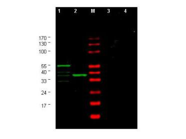
Western blot using Rocklands Affinity Purified anti-Yeast CHK1 antibody shows detection of a bands corresponding to CHK1 in Saccharomyces cerevisiae lysates. Two strains of S.cerevisiae were tested. Lane 1 shows a predominant band at ~60 kDa. Lane 2 shows a predominant band at ~38 kDa. Specific band staining is blocked when antibody is preincubated for 45 min at room temperature with 50 µg of peptide immunogen (lanes 3 and 4 respectively). Lysates were separated by 4-20% SDS-PAGE and transferred onto nitrocellulose. After blocking, the membrane was probed for 2 h at room temperature with the primary antibody diluted to 1:750 in blocking buffer diluted 1:5 in PBS. The membrane was washed and reacted with a 1:10,000 dilution of IRDye(TM)800 conjugated Gt-a-Rabbit IgG [H&L] MX (611-132-122) for 45 min at room temperature (800 nm channel, green). Molecular weight estimation was made by comparison to prestained MW markers in lane M (700 nm channel, red). IRDye(TM)800 fluorescence image was captured using the Odyssey Infrared Imaging System developed by LI-COR. IRDye is a trademark of LI-COR, Inc. Other detection systems will yield similar results.
* Mehrwertsteuer und Versandkosten nicht enthalten. Irrtümer und Preisänderungen vorbehalten