This affinity purified antibody was prepared from whole rabbit serum produced by repeated immunizations with a synthetic peptide corresponding to the N-Terminal region near amino acids 1-25 of mouse Ankrd26 protein.
0.02 M Potassium Phosphate, 0.15 M Sodium Chloride, pH 7.2
Formulierung:
Liquid (sterile filtered)
Target-Kategorie:
Mouse
Antibody Type:
Primary Antibody
Application Verdünnung:
ELISA: 1:20,000 - 1:80,000, WB: 1:500 - 1:3,000
Anwendungsbeschreibung:
This affinity purified antibody has been tested for use in ELISA and by western blot. Specific conditions for reactivity should be optimized by the end user. Expect a band approximately 81 kDa in size corresponding to Ankrd26 by western blotting in the a
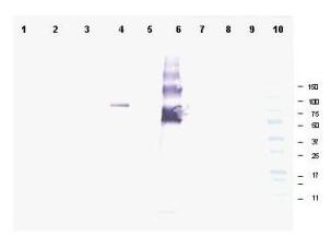
Western blot using Rocklands affinity purified anti-Ankrd26 antibody shows detection of a band at ~81 kDa corresponding to mouse Ankrd26 protein. Lane 1 Blank, Lane 2 MES cell lysate - 80 µg, Lane 3 MES cell lysate - 40 µg, Lane 4 293T-ANKRD26 transfected cell lysate - 20 µg, Lane 5 control 293T cell lysate - 20 µg, Lane 6 BSA-ANKRD26 conjugate 20 ng, Lane 7 BSA - 500 ng, Lane 8 BSA - 100 ng, Lane 9 BSA 20 ng and Lane 10 Protein standards. Detection of endogenous Ankrd26 protein in MES cell lysates may occur when detection methods with higher sensitivity are used. Proteins were separated by SDS-PAGE, transferred to nitrocellulose, and probed with the primary antibody diluted to 1:1,000 followed by detection using ALP conjugated Gt-a-Rabbit IgG (611-105-122 is suggested) diluted to 1:3,000. Size estimation was made by comparison to prestained MW markers as indicated. Personal Communication. Ira Pastan, NIH, CCR, Bethesda, MD.
* Mehrwertsteuer und Versandkosten nicht enthalten. Irrtümer und Preisänderungen vorbehalten