Anti-SMAD3 pS423pS425 antibody was prepared from whole rabbit serum produced by repeated immunizations with a dual phosphorylated synthetic peptide corresponding to a c-terminal region with Serine 423 and Serine 425 of human SMAD3 protein.
Konjugation:
Unconjugated
Alternative Synonym:
rabbit anti-SMAD3 pS423pS425 antibody, SMAD-3, SMAD 3, mothers against decapentaplegic homolog 3 antibody, MAD homolog 3, Mothers against DPP homolog 3, SMAD family member 3, MADH3, MADH 3, JV15-2
This affinity purified antibody has been tested for use in ELISA, immunohistochemistry and by western blot. Specific conditions for reactivity should be optimized by the end user. Expect a band approximately 48 kDa in size corresponding to phosphorylated
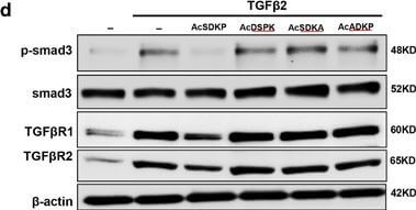
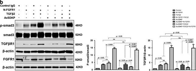
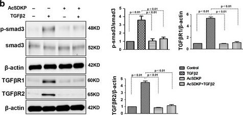
Proximity between AcSDKP and FGFR1 inhibits the TGFbeta/smad signaling pathway in HMVECs. (a) HMVECs were treated with N-FGFR1 (1.5µg/ml) for 48h with or without preincubation with AcSDKP (100nM) for 2h, and the proximity between AcSDKP and FGFR1 was analyzed by the Duolink In Situ Assay. For each slide, images at a * 400 original magnification were obtained from six different areas. (b and c) HMVECs were treated with TGFbeta2 (5ng/ml) for 15min or 48h with or without preincubation with AcSDKP for 2h, and the p-smad3, TGFbetaR1, TGFbetaR2 and FGFR1 levels were analyzed by western blot. Densitometric analysis of the p-smad3/smad3, TGFbetaR1/beta-actin, TGFbetaR2/beta-actin and FGFR1/beta-actin levels from each group (n=6) were analyzed. (d and e) HMVECs were incubated with TGFbeta2 for 15min or 48h with or without preincubation with AcSDKP or its mutants (AcDSPK, AcSDKA, AcADKP) (100nM) for 2h. The p-smad3/smad3, TGFbetaR1/beta-actin, TGFbetaR2/beta-actin and FGFR1/beta-actin protein levels were analyzed by western blot Figure provided by CiteAb. Source: Cell Death Dis, PMID: 28
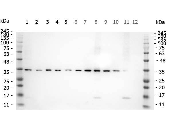
Western Blot of Rabbit anti-SMAD3 pS423 pS425 antibody. Marker: Opal Pre-stained ladder (p/n MB-210-0500). Lane 1: HEK293 lysate (p/n W09-000-365). Lane 2: HeLa Lysate (p/n W09-000-364). Lane 3: MCF-7 Lysate (p/n W09-000-360). Lane 4: Jurkat Lysate (p/n W09-000-370). Lane 5: A431 Lysate (p/n W09-000-361). Lane 6: A549 Lysate (p/n W09-001-372). Lane 7: LNCap Lysate (p/n W09-001-GJ9). Lane 8: MOLT-4 Lysate (p/n W09-001-GK2). Lane 9: Ramos Lysate (p/n W09-000-GK4). Lane 10: Raji Lysate (p/n W09-001-368). Lane 11: A-172 Lysate (p/n W09-001-GL5). Lane 12: NIH/3T3 Lysate (p/n W10-000-358). Load: 10 µg per lane. Primary antibody: SMAD3 pS423 pS425antibody at 1ug/mL overnight at 4C. Secondary antibody: Peroxidase rabbit secondary antibody (p/n 611-103-122) at 1:30,000 for 60 min at RT. Blocking Buffer: 1% Casein-TTBS (p/n MB-082) for 30 min at RT. Predicted/Observed size: 35 kDa for SMAD3 pS423 pS425.
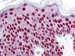
Rocklands affinity purified anti-Smad3 pS423 pS425 antibody was used at 2.5 ug/ml to detect signal in a variety of tissues including multi-human, multi-brain and multi-cancer slides. This image shows strong nuclear staining in the majority of epidermal keratinocytes at 40X. Tissue was formalin-fixed and paraffin embedded. The image shows localization of the antibody as the precipitated red signal, with a hematoxylin purple nuclear counterstain. Personal Communi-cation, Tina Roush, LifeSpanBiosciences, Seattle, WA.
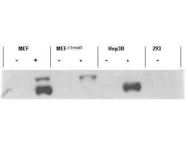
Western blot using Rocklands affinity purified anti-Smad3 pS423 pS425 antibody shows detection of endogenous Smad3 in stimulated cell lysates. Lysates were prepared from control cells (- lanes), or cells stimulated with 2 ng/ml TGF (+lanes) for 1 hour. This reagent recognizes phosphorylated Smad3 and has negligible reactivity against non-phosphorylated Smad3 protein. Personal Communication. Ying Zhang, NIH, CCR, Bethesda, MD.
Proximity between AcSDKP and FGFR1 inhibits the TGFbeta/smad signaling pathway in HMVECs. (a) HMVECs were treated with N-FGFR1 (1.5µg/ml) for 48h with or without preincubation with AcSDKP (100nM) for 2h, and the proximity between AcSDKP and FGFR1 was analyzed by the Duolink In Situ Assay. For each slide, images at a * 400 original magnification were obtained from six different areas. (b and c) HMVECs were treated with TGFbeta2 (5ng/ml) for 15min or 48h with or without preincubation with AcSDKP for 2h, and the p-smad3, TGFbetaR1, TGFbetaR2 and FGFR1 levels were analyzed by western blot. Densitometric analysis of the p-smad3/smad3, TGFbetaR1/beta-actin, TGFbetaR2/beta-actin and FGFR1/beta-actin levels from each group (n=6) were analyzed. (d and e) HMVECs were incubated with TGFbeta2 for 15min or 48h with or without preincubation with AcSDKP or its mutants (AcDSPK, AcSDKA, AcADKP) (100nM) for 2h. The p-smad3/smad3, TGFbetaR1/beta-actin, TGFbetaR2/beta-actin and FGFR1/beta-actin protein levels were analyzed by western blot Figure provided by CiteAb. Source: Cell Death Dis, PMID: 28771231.
* Mehrwertsteuer und Versandkosten nicht enthalten. Irrtümer und Preisänderungen vorbehalten