This affinity purified antibody was prepared from whole rabbit serum produced by repeated immunizations with a synthetic peptide corresponding to amino acids near the carboxyl terminus of human TAF1.
0.02 M Potassium Phosphate, 0.15 M Sodium Chloride, pH 7.2
Formulierung:
Liquid (sterile filtered)
Target-Kategorie:
Human
Antibody Type:
Primary Antibody
Application Verdünnung:
ELISA: 1:150,000 - 1:300,000, ChIP: User Optimized, IHC: 10µg/ml, IP: User Optimized, WB: 1:100 - 1:2,000
Anwendungsbeschreibung:
This affinity purified antibody has been tested for use in ELISA, Immunohistochemistry, ChIP, and western blotting. Specific conditions for reactivity should be optimized by the end user. Expect a band approximately 213 kDa in size corresponding to TAF1
Transcription Initiation Factor TFIID Subunit 1 (TAF1) antibody was used to detect TAF1 in treated and untreated HeLa Cells. HeLa cells were treated with TNF alpha and Chromatin was prepared by EZ Magna Chip Kit (Millipore). CHIP was performed on fos promoters using 5 µg of TAF1 antibody from Rockland (P/n 600-401-995) and an RNA PolII antibody. Image with data provided courtesy of Shiraz Mujtaba, Ph.D., Dept. of Structural & Chemical Biology, Mount Sinai School of Medicine.
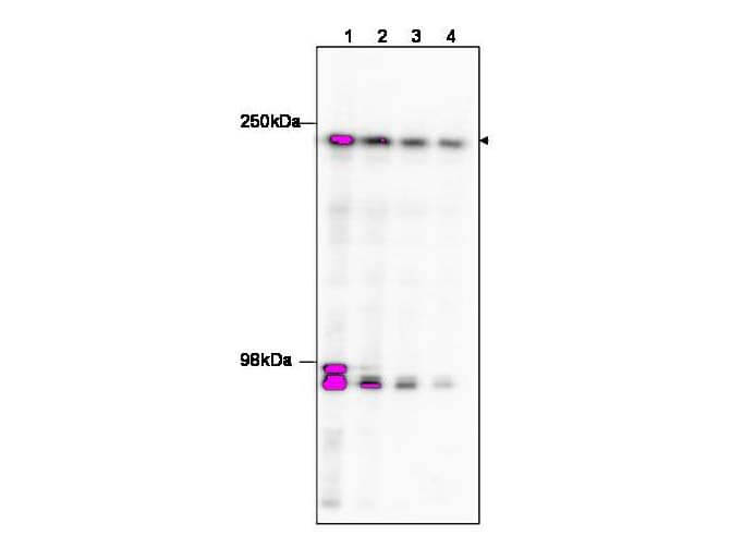
Western blot using Rocklands affinity purified anti-TAF1 to detect TAF1 in HeLa nuclear extract (arrowhead). The membrane was probed with the primary antibody at dilutions of 1:100 (lane 1), 1:250 (lane 2), 1: 500 (Lane 3 and 1:1,000 (Lane 4). The identity of the bands at ~95 kDa is unknown, but may be degraded TAF1. Personal Communication, Anne Gegonne, CCR-NCI, Bethesda, MD.
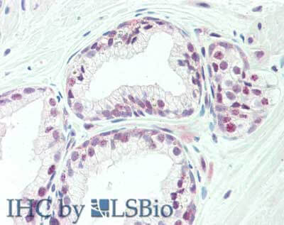
Immunohistochemistry of rabbit anti-TAF1 antibody. Tissue: prostate. Fixation: formalin fixed paraffin embedded. Antigen retrieval: not required. Primary antibody: Anti-TAF1 at 10 µg/mL for 1 h at RT. Secondary antibody: Peroxidase rabbit secondary antibody at 1:10,000 for 45 min at RT. Staining: TAF-1 as precipitated red signal with hematoxylin purple nuclear counterstain.
Western blot using Rocklands affinity purified anti-TAF1 to detect TAF1 in HeLa nuclear extract (arrowhead). The membrane was probed with the primary antibody at dilutions of 1:100 (lane 1), 1:250 (lane 2), 1: 500 (Lane 3 and 1:1,000 (Lane 4). The identity of the bands at ~95 kDa is unknown, but may be degraded TAF1. Personal Communication, Anne Gegonne, CCR-NCI, Bethesda, MD.
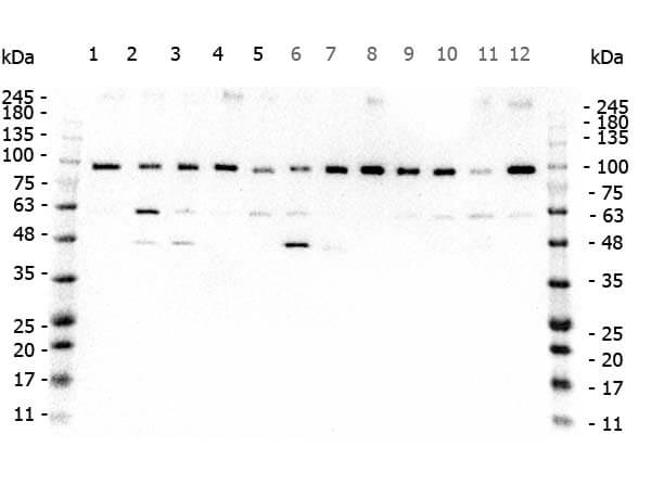
Western Blot of Rabbit anti-TAF1 antibody. Marker: Opal Pre-stained ladder (p/n MB-210-0500). Lane 1: HEK293 lysate (p/n W09-000-365). Lane 2: HeLa Lysate (p/n W09-000-363). Lane 3: MCF-7 Lysate (p/n W09-000-360). Lane 4: Jurkat Lysate (p/n W09-000-370). Lane 5: A431 Lysate (p/n W09-000-361). Lane 6: A549 Lysate (p/n W09-001-372). Lane 7: LNCap Lysate (p/n W09-001-GJ9). Lane 8: MOLT-4 Lysate (p/n W09-001-GK2). Lane 9: Ramos Lysate (p/n W09-000-GK4). Lane 10: Raji Lsyate (p/n W09-001-368). Lane 11: A-172 Lysate (p/n W09-001-GL5). Lane 12: NIH/3T3 Lysate (p/n W10-000-358). Load: 35 µg per lane. Primary antibody: TAF1 antibody at 0.2ug/mL overnight at 4C. Secondary antibody: Peroxidase rabbit secondary antibody (p/n 611-103-122) at 1:30,000 for 60 min at RT. Blocking Buffer: 1% Casein-TTBS for 30 min at RT. Predicted/Observed size: 250kDa for TAF1.
* Mehrwertsteuer und Versandkosten nicht enthalten. Irrtümer und Preisänderungen vorbehalten