This affinity purified antibody was prepared from whole rabbit serum produced by repeated immunizations with a peptide corresponding to amino acids near the N-terminal end of human PTEN-P1 protein.
Konjugation:
Unconjugated
Alternative Synonym:
rabbit anti-PTEN-P1 antibody, Phosphatase and tensin homolog 2, Phosphatidylinositol 3,4,5-trisphosphate 3-phosphatase and dual-specificity protein phosphatase PTEN, Mutated in multiple advanced cancers 1, Phosphatase and tensin homolog, PTEN, MMAC1, TEP1
0.02 M Potassium Phosphate, 0.15 M Sodium Chloride, pH 7.2
Formulierung:
Liquid (sterile filtered)
Target-Kategorie:
Human
Antibody Type:
Primary Antibody
Application Verdünnung:
ELISA: 1:160,000, WB: 1:500 to 1:2,000
Anwendungsbeschreibung:
This affinity purified antibody has been tested for use in ELISA and western blot. Specific conditions for reactivity should be optimized by the end user. Expect a band approximately 55 kDa in size corresponding to PTEN-P1 protein by western blotting in
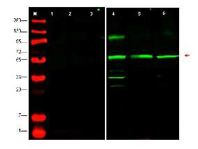
Western blot using Rocklands affinity purified anti-PTEN-P1 antibody shows detection at ~55kDa (arrowhead) of endogenous PTEN-P1 in whole cell lysates from human derived cell lines HeLa (p/n W09-000-364) [lane 4], HEK293 (p/n W09-000-365) [lane 5] and MCF7 (p/n W09-000-360) [lane 6]. Lanes 1-3 were show the results of staining after the antibody was first pre-incubated with the immunizing peptide. The identity of lower molecular weight bands in lane 4 is unknown. Briefly, each lane contains approximately 35µg of lysate. Primary antibody was used at a 1:500 dilution in 5% BLOTTO in PBS reacted overnight at 4C. The membrane was washed and reacted with a 1:10,000 dilution of IRDye800(TM) conjugated Gt-a-Rabbit IgG [H&L] MX (p/n 611-132-122) for 45 min at room temperature (800 nm channel, green). Molecular weight estimation was made by comparison to prestained MW markers in lane M (700 nm channel, red). IRDye(TM)800 fluorescence image was captured using the Odyssey Infrared Imaging System developed by LI-COR. IRDye is a trademark of LI-COR, Inc. Other detection systems will yield similar results.
* Mehrwertsteuer und Versandkosten nicht enthalten. Irrtümer und Preisänderungen vorbehalten