This affinity purified antibody was prepared from whole rabbit serum produced by repeated immunizations with a synthetic peptide corresponding to an internal region of Human Casz1 protein.
Konjugation:
Unconjugated
Alternative Synonym:
rabbit anti-CASZ1 Antibody, CASZ1, Zinc finger protein castor homolog 1, Castor-related protein, Zinc finger protein 693, CST, SRG, ZNF693
0.02 M Potassium Phosphate, 0.15 M Sodium Chloride, pH 7.2
Formulierung:
Liquid (sterile filtered)
Target-Kategorie:
Human
Antibody Type:
Primary Antibody
Application Verdünnung:
ELISA: 1:45,000, IHC: User Optimized, IF Microscopy: User Optimized, WB: 1:1,000 - 1:5,000
Anwendungsbeschreibung:
This antibody has been tested for use in ELISA, IHC, IF, and western blotting. This antiserum detects endogenous Casz1 proteins, both the hCasz5 and hCasz11 isoforms. Expect a band approximately 125 kDa for hCasz5 and 190 kDa for hCaz11 in size correspon
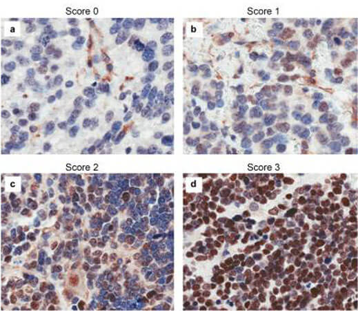
Immunohistochemistry results of Rabbit Anti-hCASZ1 Antibody. Tissue: NB patient tumor. A. Score 0- a rare positive nuclei. B. Score 1- (1-10% positive) equivocal/uninterpretable. C. Score 2- (10-50% positive) weak positive. D. Score 3- (>50% positive) strong positive. Primary Antibody: Rabbit Anti-CASZ1 stained brown. Nucleus counterstained with hematoxylin (blue). Localization: Nuclear.
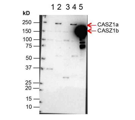
Western Blot of Anti-CASZ1 Antibody. Lane 1: NBLS Cytoplasmic (20µg). Lane 2: NBLS Nuclear (3µg). Lane 3: BE2C Cytoplasmic (30µg). Lane 4: BE2C Nuclear (7µg). Lane 5: SY5Y-CASZ1b (10µg). Block: 5% Blotto/TTBS for 1 hour. Primary: Casz1 1:10,000 for 1 hour. Secondary: Goat anti-Rabbit HRP for 1 hour. 240sec exposure. Detects nuclear endogenous CASZ1a and CASZ1b, and transiently transfected CASZ1b isoform. Personal communication and images from Carol Thiele Galetto, NCI.
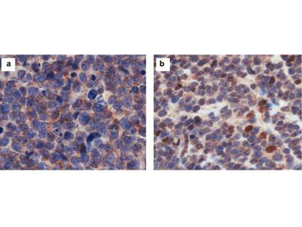
Immunohistochemistry results of Rabbit Anti-hCasz1 Antibody. Tissue: NB patient tumor. A. CASZ1 localized exclusively in the cytoplasm. B. CASZ1 localized in the cytoplasm and nucleus. Primary Antibody: Rabbit Anti-CASZ1 stained brown. Nucleus counterstained with hematoxylin (blue).
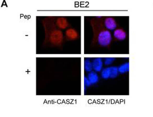
Immunofluorescence results of Endogenous CASZ1. Cells: BE2 cells. With or without Pre-Incubation of Anti-CASZ1 Antibody with CASZ1 Peptide. Staining: Rabbit Anti-CASZ1 Antibody. Chromatin counter stain: DAPI.
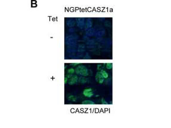
Immunofluorescence results of Rabbit Anti-CASZ1 Antibody. Tissue: Mouse Xenograft tumor of human NB cell line transfected with or without tetracycline inducible CASZ1 (NGPtetCASZ1a). Antibody: Rabbit Anti-CASZ1 Antibody. Counterstain: DAPI.
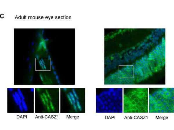
Immunofluorescence of Rabbit anti-CASZ1 Antibody. Tissue: adult murine ocular tissue. Antibody: Rabbit Anti-CASZ1 Antibody. Counterstain: DAPI. Localization: nucleus in lens epithelia but primarily localizes in the cytoplasm in photoreceptor cells.
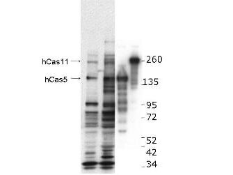
Western blot using Rocklands anti-hCASZ1 antibody. This blot shows detection of endogenous and transfected human CASZ1 protein in fresh whole cell lysate (~30 µg). Lane 1: BE2(s) cell lysate. Lane 2: BE2(N) cell lysate. Lane 3: SY5Y transfected with hCASZ5 (125kDa). Lane 4: SY5Y transfected with hCASZ11 (190kDa). Protein was resolved by SDS-PAGE and transferred onto nitrocellulose. After blocking, the membrane was probed with the primary antibody diluted to 1:1,000 for 1.5 hours at room temperature then incubated with HRP-conjugated Goat Anti-Rabbit antibody for 45 min. at room temperature. Personal communication, Carol Thiele, NCI, Bethesda, MD.
* Mehrwertsteuer und Versandkosten nicht enthalten. Irrtümer und Preisänderungen vorbehalten