Anti-PKR antibody was prepared from whole rabbit serum produced by repeated immunizations with a peptide corresponding to 14 amino acids near the C-terminus of human PKR. The immunogen is located within the last 50 amino acids of PKR.
Konjugation:
Unconjugated
Alternative Synonym:
PKR Antibody, PKR, PRKR, EIF2AK1, PKR, Interferon-induced, double-stranded RNA-activated protein kinase, Eukaryotic translation initiation factor 2-alpha kinase 2, eIF-2A protein kinase 2
Anti-PKR Antibody has been tested for use in ELISA, Western Blotting, Immunohistochemistry, and Immunofluorescence. Specific conditions for reactivity should be optimized by the end user. Expect a band at approximately 62 kDa in Western Blots of specific
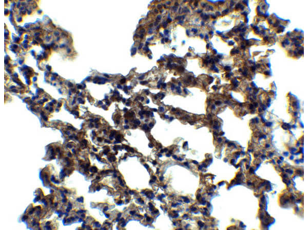
Immunohistochemistry of Rabbit Anti-PKR Antibody. Tissue: Mouse Lung. Fixation: paraffin-embedded formaldehyde and blocked with 10% serum for 1 h at RT. Antigen retrieval: heat mediation with a citrate buffer (pH6). Primary Antibody: anti-PKR antibody at 5µg/ml overnight at 4C. Secondary Antibody: goat anti-rabbit IgG H&L (HRP) at 1/250. Counter stained with Hematoxylin.
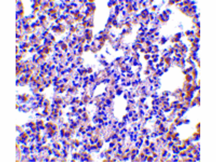
Immunohistochemistry of PKR antibody. Tissue: rat lung tissue. Fixation: formalin fixed paraffin embedded. Antigen retrieval: not required. Primary antibody: PKR antibody at 2.5 µg/mL for 1 h at RT. Secondary antibody: Peroxidase rabbit secondary antibody at 1:10,000 for 45 min at RT. Localization: PKR is nuclear and cytoplasmic. Staining: PKR is stained with toluidine blue.
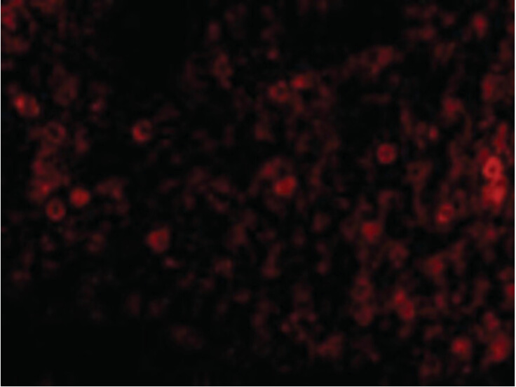
Immunofluorescence Microscopy of PKR antibody. Cell Type: rat lung cells. Fixation: 0.5% PFA. Antigen retrieval: not required. Primary antibody: PKR antibody at 20 µg/mL for 1 h at RT. Secondary antibody: Fluorescein rabbit secondary antibody at 1:10,000 for 45 min at RT. Localization: PKR is nuclear and cytoplasmic. Staining: PKR as red fluorescent signal.
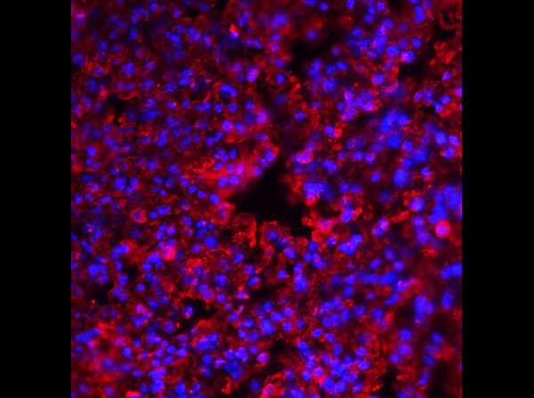
Immunofluorescence of Rabbit Anti-PKR Antibody. Tissue: Mouse Lung. Fixation: 4% paraformaldehyde-fixed Primary Antibody: PKR at 20µg/mL, Secondary Antibody: goat anti-rabbit IgG antibody at 1/500 dilution (red) and DAPI staining (blue).
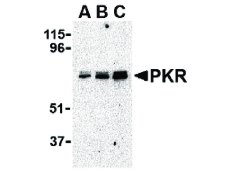
Western Blot of PKR antibody in A431 whole cell lysate. Lane A: PKR antibody at 0.5 µg/mL. Lane B: PKR antibody at 1 µg/mL. Lane C: PKR antibody at 2 µg/mL. Load: 35 µg per lane. Primary antibody: PKR antibody at designated concentrations for overnight at 4C. Secondary antibody: Peroxidase rabbit secondary antibody at 1:10,000 for 45 min at RT. Block: 5% BLOTTO overnight at 4C. Predicted/Observed size: 62 kDa, 72 kDa for PKR.
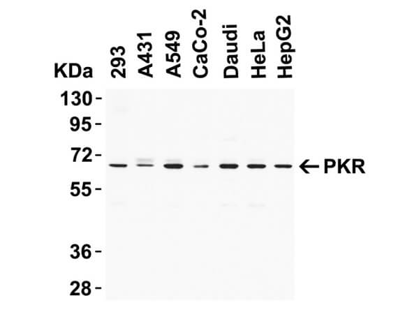
Western Blot of Rabbit Anti-PKR Antibody. Lysates: Human Cell Lines. Loading: 15µg of lysates per lane. Primary Antibody: PKR (1µg/mL), 1hr at RT in 5% NFDM/TBST. Secondary Antibody: Goat anti-rabbit IgG HRP conjugate at 1:10,000. Expect: ~62kDa.
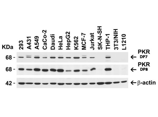
Western Blot of Anti-PKR Antibody. Loading: 15 µg of lysates per lane. Primary Antibodies: PKR (p/n 600-401-DP7) (1 µg/mL), PKR (p/n 600-401-DP8) (1 µg/mL), and beta-actin (1 µg/mL), 1hr at RT in 5% NFDM/TBST. Secondary Antibody: Goat anti-rabbit IgG HRP conjugate at 1:10,000. Expect: ~62kDa.
* Mehrwertsteuer und Versandkosten nicht enthalten. Irrtümer und Preisänderungen vorbehalten