Anti-TIM-1 antibody was prepared from whole rabbit serum produced by repeated immunizations with a 16 amino acid synthetic peptide from near the N-terminus of human TIM-1.
Konjugation:
Unconjugated
Alternative Synonym:
TIM-1 Antibody, TIM, KIM1, TIM1, HAVCR, KIM-1, TIMD1, TIMD-1, HAVCR-1, Hepatitis A virus cellular receptor 1, Kidney injury molecule 1, HAVcr-1, T-cell immunoglobulin and mucin domain-containing protein 1, T-cell immunoglobulin mucin receptor 1, T-cell membrane protein 1, CD365
Anti-TIM-1 Antibody has been tested for use in ELISA, Western Blotting, Immunohistochemistry and Immunofluorescence. Specific conditions for reactivity should be optimized by the end user. Expect a band at approximately 39 kDa in Western Blots of specifi
Immunofluorescence Microscopy of TIM-1 antibody. Cell Type: human uterus cells. Fixation: 0.5% PFA. Antigen retrieval: not required. Primary antibody: TIM-1 antibody at 20 µg/mL for 1 h at RT. Secondary antibody: Fluorescein rabbit secondary antibody at 1:10,000 for 45 min at RT. Localization: TIM-1 is nuclear and chromosomal. Staining: TIM-1 as red fluorescent signal.
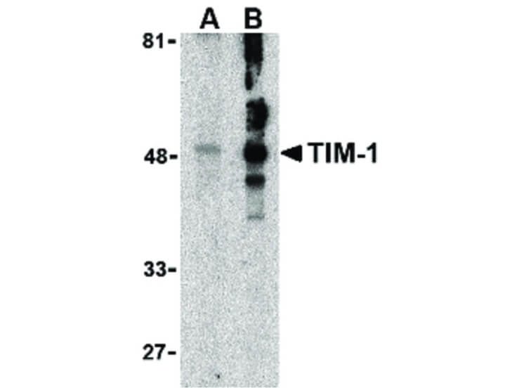
Western Blot of TIM-1 antibody in human uterus tissue lysate. Lane A: TIM-1 antibody at 1 µg/mL. Lane B: TIM-1 antibody at 2 µg/mL. Load: 35 µg per lane. Primary antibody: TIM-1 antibody at designated concentrations for overnight at 4C. Secondary antibody: Peroxidase rabbit secondary antibody at 1:10,000 for 45 min at RT. Block: 5% BLOTTO overnight at 4C. Predicted/Observed size: 75 kDa, 48 kDa for TIM-1. Other band(s): TIM-1 splice variants and isoforms.
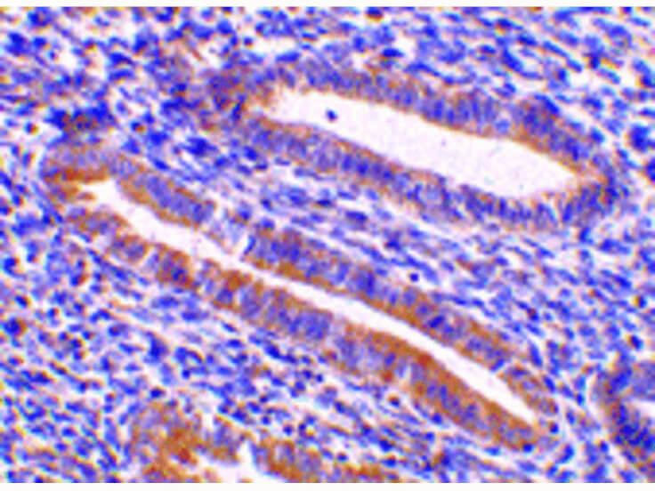
Immunohistochemistry of TIM-1 antibody. Tissue: human uterus tissue. Fixation: formalin fixed paraffin embedded. Antigen retrieval: not required. Primary antibody: TIM-1 antibody at 10 µg/mL for 1 h at RT. Secondary antibody: Peroxidase rabbit secondary antibody at 1:10,000 for 45 min at RT. Localization: TIM-1 is nuclear and chromosomal. Staining: TIM-1 is stained with toluidine blue.
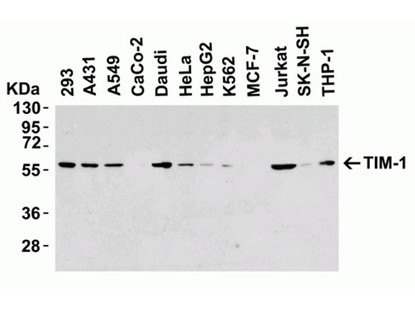
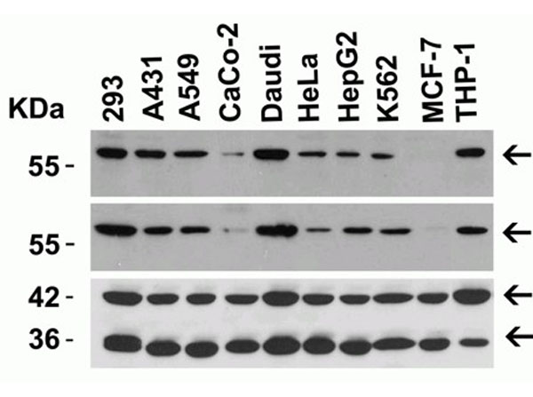
Western Blot of TIM-1. Load: 15 µg of human cell lysates per lane. Primary Antibody: TIM-1 at 8 µg/mL for overnight incubation at 4 C in 5% NFDM/TBST. Secondary: Goat anti-rabbit IgG HRP conjugate at 1:10000 dilution.
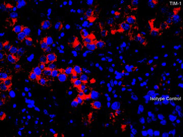
Immunofluorescence of TIM-1. Tissue: Human Testis. Fixation: 4% paraformaldehyde-fixed. Primary Antibody: TIM-1 at 10 µg/mL. Secondary: goat anti-rabbit IgG secondary antibody at 1:500 dilution (red) and DAPI staining (blue).
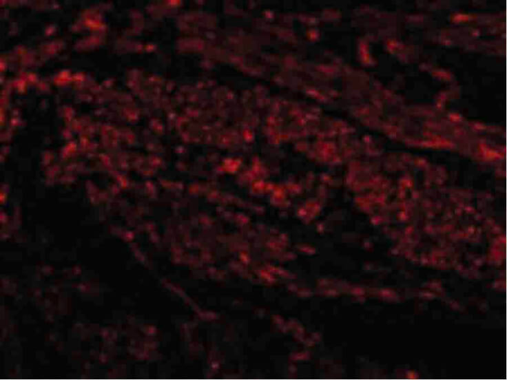
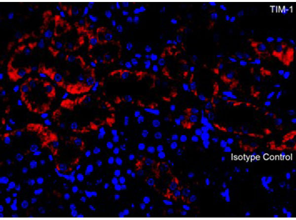
Immunofluorescence of TIM-1.Tissue: Mouse Kidney. Fixation: 4% paraformaldehyde-fixed. Primary Antibody: TIM-1 at 10 µg/mL. Secondary: goat anti-rabbit IgG secondary antibody at 1:500 dilution (red) and DAPI staining (blue).
Western Blot of TIM-1. Load: 15 µg of lysates per lane. Primary Antibody: Top: TIM-1 (p/n 600-401-FE6) at 8 µg/mL, Middle: TIM1 at 1 µg/mL for overnight incubation at 4 C, Bottom: beta-actin at 1 µg/mL and GAPDH at 0.02 µg/mL for 1h incubation at RT, in 5% NFDM/TBST. Secondary: Goat anti-rabbit IgG HRP conjugate at 1:10000 dilution.
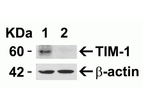
Western Blot of TIM-1. Load: 15 µg of cell lysates per lane. Lane 1: HeLa cells were transfected with control siRNAs, Lane 2: TIM-1 siRNA Knockdown in Hela cells. Primary Antibody: TIM-1 at 8 µg/mL for 1 h incubation at RT in 5% NFDM/TBST. Secondary: Goat anti-rabbit IgG HRP conjugate at 1:10000 dilution.
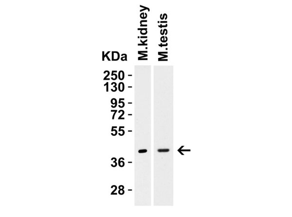
Western Blot of TIM-1. Load: 15 µg of mouse tissue lysates. Primary Antibody: TIM-1 at 2 µg/mL for 1h incubation at RT in 5% NFDM/TBST. Secondary: Goat anti-rabbit IgG HRP conjugate at 1:10000 dilution.
* Mehrwertsteuer und Versandkosten nicht enthalten. Irrtümer und Preisänderungen vorbehalten