CX3CR1 Antibody was produced from whole rabbit serum prepared by repeated immunizations with a peptide corresponding to an internal region of human CX3CR1. The sequence differs from those of mouse and rat CX3CR1 by four amino acids.
0.02 M Potassium Phosphate, 0.15 M Sodium Chloride, pH 7.2
Formulierung:
Liquid (sterile filtered)
Target-Kategorie:
Human
Antibody Type:
Primary Antibody
Application Verdünnung:
Flow Cytometry: 0.1µg/mL, IHC: 2µg/mL, IF Microscopy: 10-20µg/mL, WB: 1-2µg/mL
Anwendungsbeschreibung:
Anti-CX3CR1 Antibody is tested for use in E, WB, IHC, IF and FACS. Expect a band approximately ~40.3 kDa on specific lysates. Western Blot in human, mouse and rat samples, Immunohistochemistry in rat samples, Immunofluorescence in human, mouse and rat sa
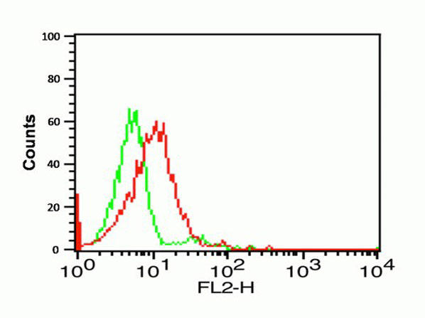
Flow Cytometry Validation of CX3CR1. Cells: THP-1 Cells. Overlay histogram showing THP-1 cells stained with Anti-CX3CR1 at 1µg/1x106cells (red line) for 1h at 4C in 2% FBS/PBS. Followed by secondary antibody 488 goat anti-rabbit IgG (H+L) at 1:500 dilution for 1h at 4C. Isotype control rabbit IgG1 antibody at 1µg/1x106cells (green line) used under the same conditions. Acquisition of >10,000 events was performed.
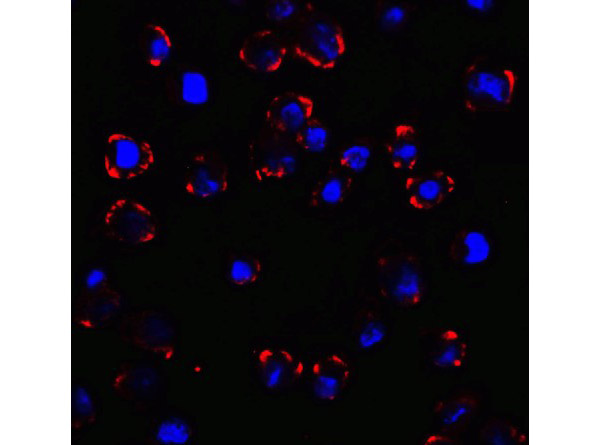
Immunofluorescence Validation of CX3CR1. Cells: K562 cells. Fixation: 4% paraformaldehyde-fixed. Primary Antibody: CX3CR1 at 10 µg/mL. Secondary: goat anti-rabbit IgG secondary antibody at 1:500 dilution (red) and DAPI staining (blue). Image showing membrane staining on K562 cells.
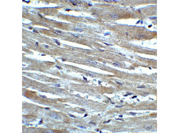
Immunohistochemistry Validation of CX3CR1. Tissue: Rat Heart Tissue. Fixation: paraffin-embedded, fixed with formaldehyde and blocked with 10% serum for 1 h at RT. Antigen retrieval: heat mediation with a citrate buffer (pH6). Primary Antibody: anti-CX3CR1 antibody at 2 µg/ml overnight at 4C. Secondary: goat anti-rabbit IgG H&L (HRP) at 1:250. Counter stained with Hematoxylin.
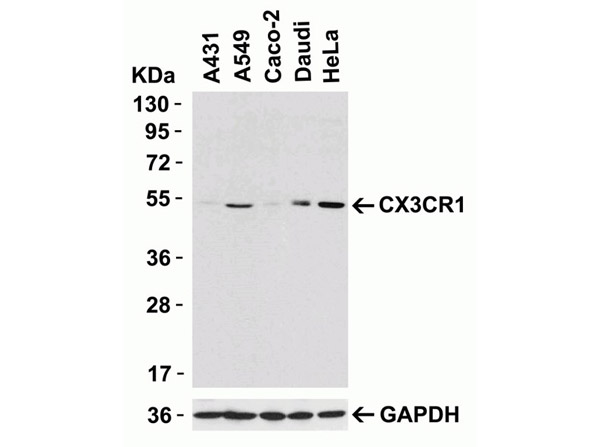
Western Blot Validation of Anti-CX3CR1.Loading: 15 µg of human lysates per lane. Lane 1: A431, Lane 2: A549, Lane 3: Caco-2, Lane 4: Daudi, Lane 5: HeLa. Primary Antibody: Anti-CX3CR1 at 0.5µg/mL for 1h at RT in 5% NFDM/TBST. Secondary: Goat anti-rabbit IgG HRP conjugate at 1:10000 dilution.
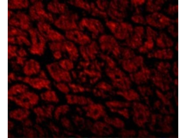
Immunofluorescence Validation of CX3CR1. Tissue: Human Heart. Fixation: 4% paraformaldehyde-fixed. Primary Antibody: CX3CR1 at 10 µg/mL. Secondary: goat anti-rabbit IgG secondary antibody at 1/500 dilution (red).
KD Validation Western Blot of CX3CR1. Loading: 15 µg of lysates per lane. Lane 1: 293 cells transfected with control siRNAs. Lane 2: 293 cells transfected with CX3CR1 siRNAs. Primary Antibody: Anti-CX3CR1 at 0.5µg/mL for 1h at RT in 5% NFDM/TBST. Secondary: Goat anti-rabbit IgG HRP conjugate at 1:10000 dilution.
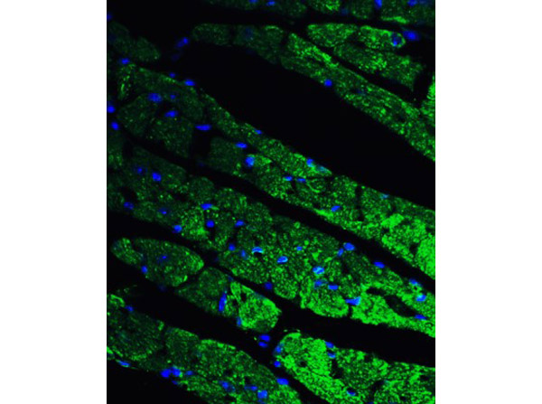
Immunofluorescence Validation of CX3CR1. Tissue: Mouse Heart Tissue. Fixation: 4% paraformaldehyde-fixed. Primary Antibody: CX3CR1 at 20 µg/mL. Secondary: goat anti-rabbit IgG secondary antibody at 1:500 dilution (green) and DAPI staining (blue).
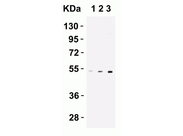
Western Blot Validation of Anti-CX3CR1. Loading: 15 µg of THP1 cell lysates per lane. Primary Antibody: Anti-CX3CR1 at (Lane 1: 0.2 µg/mL Lane 2: 0.5 µg/mL Lane 3: 1 µg/mL) for 1h at RT in 5% NFDM/TBST. Secondary: Goat anti-rabbit IgG HRP conjugate at 1:10000 dilution.
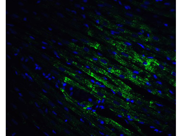
Immunofluorescence Validation of CX3CR1. Tissue: Rat Heart Tissue. Fixation: 4% paraformaldehyde-fixed. Primary Antibody: CX3CR1 at 20 µg/mL.Secondary: goat anti-rabbit IgG secondary antibody at 1:500 dilution (green) and DAPI staining (blue).
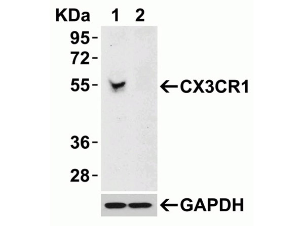
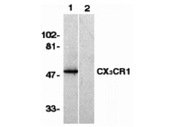
Western Blot Validation of Anti-CX3CR1. Loading: 15 µg of Human Spleen lysates per lane. Primary Antibody: Anti-CX3CR1 at 1µg/mL in the absence (lane 1) or presence of blocking peptide (lane 2) for 1h at RT in 5% NFDM/TBST. Secondary: Goat anti-rabbit IgG HRP conjugate at 1:10000 dilution.
* Mehrwertsteuer und Versandkosten nicht enthalten. Irrtümer und Preisänderungen vorbehalten