CXCR4 Antibody was produced from whole rabbit serum prepared by repeated immunizations with a peptide corresponding to an internal sequence in the second extracellular loop (EL) of human CXCR4.
0.02 M Potassium Phosphate, 0.15 M Sodium Chloride, pH 7.2
Formulierung:
Liquid (sterile filtered)
Target-Kategorie:
Human
Antibody Type:
Primary Antibody
Application Verdünnung:
IHC: 5µg/mL, IF Microscopy: 4µg/mL, WB: 1-2µg/mL
Anwendungsbeschreibung:
Anti-CXCR4 Antibody is tested for use in E, IF, IHC, and WB. Expect a band approximately ~39.7 kDa on specific lysates. Specific conditions for reactivity should be optimized by the end user.
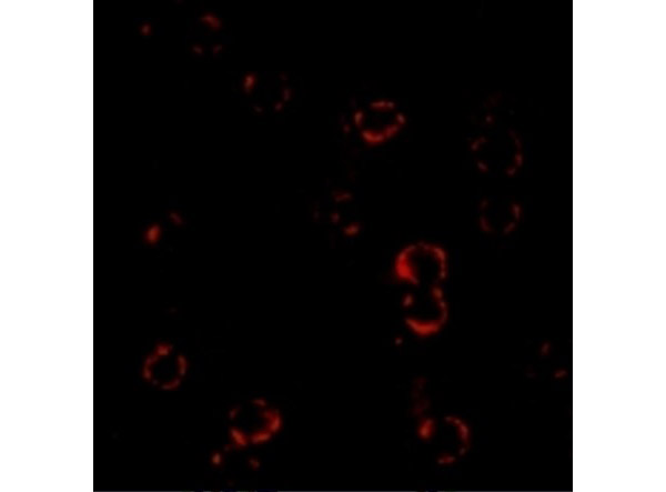
Immunofluorescence Validation of CXCR4. Cells: HeLa cells. Fixation: 4% paraformaldehyde. Primary: Anti-CXCR4 at 4 µg/mL. Secondary: goat anti-rabbit IgG at 1:500 dilution (red). Image showing both membrane and cytoplasmic staining on HeLa cells.
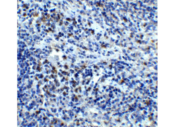
Immunohistochemistry Validation of CXCR4. Tissue: Human Spleen. Fixation: paraffin-embedded, formaldehyde and blocked with 10% serum for 1 h at RT. Antigen retrieval: heat mediation with a citrate buffer (pH6). Primary: anti-CXCR4 antibody at 5 µg/ml overnight at 4C. Secondary: goat anti-rabbit IgG H&L (HRP) at 1:250. Counter stained with Hematoxylin.
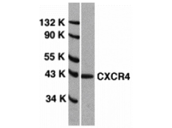
Western Blot Validation of CXCR4.Loading: 15 µg of HeLa cell lysates per lane. Primary Antibody: Anti-CXCR4 at 1µg/mL for 1h at RT in 5% NFDM/TBST. Secondary: Goat anti-rabbit IgG HRP conjugate at 1:10000 dilution.
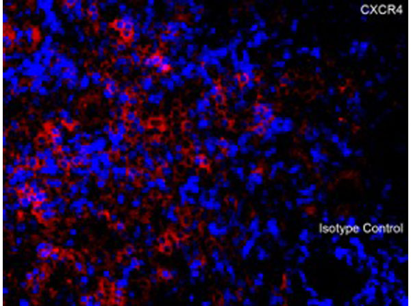
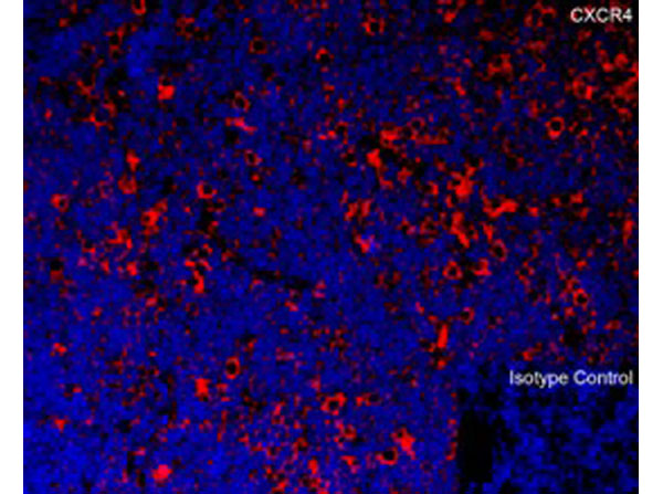
Immunofluorescence of CXCR4. Tissue: Human Spleen. Fixation: 4% paraformaldehyde-fixed. Primary Antibody: CXCR4 at 20 µg/mL. Secondary: goat anti-rabbit IgG antibody at 1:500 dilution (red) and DAPI staining (blue).
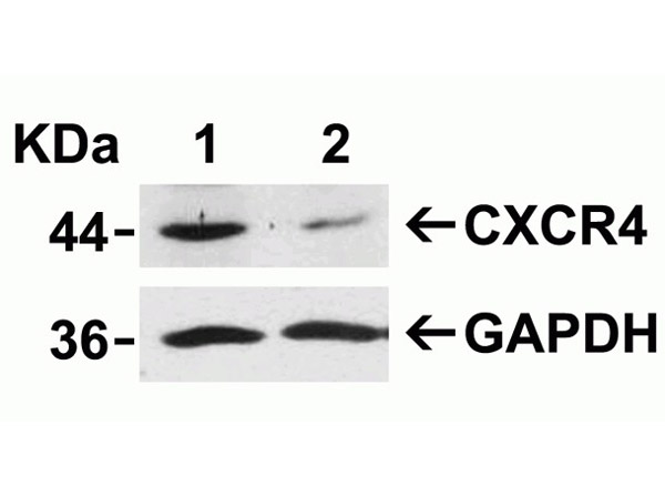
Western Blot Validation of CXCR4 siRNA Knockdown. Loading: 10 µg of HeLa whole cell lysates per lane. HeLa cells were transfected with control siRNAs (lane 1) or CXCR4 siRNAs (lane 2). Primary Antibody: Anti-CXCR4 at 2µg/mL for 1h at RT in 5% NFDM/TBST. Secondary: Goat anti-rabbit IgG HRP conjugate at 1:10000 dilution.
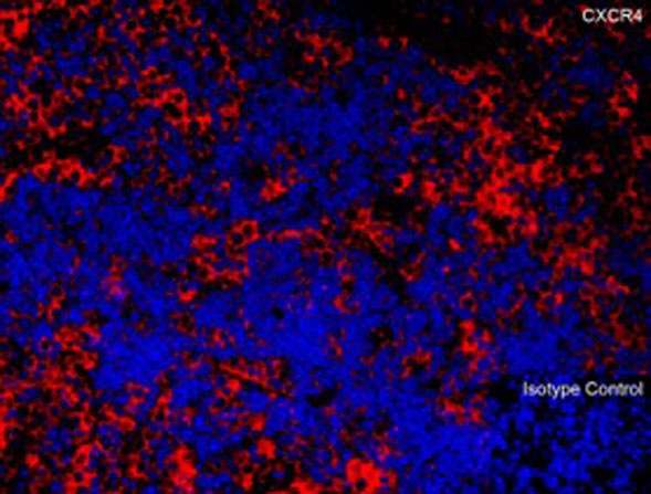
Immunofluorescence of CXCR4. Tissue: Mouse Thymus. Fixation: 4% paraformaldehyde-fixed. Primary Antibody: CXCR4 at 20 µg/mL. Secondary: goat anti-rabbit IgG antibody at 1:500 dilution (red) and DAPI staining (blue).
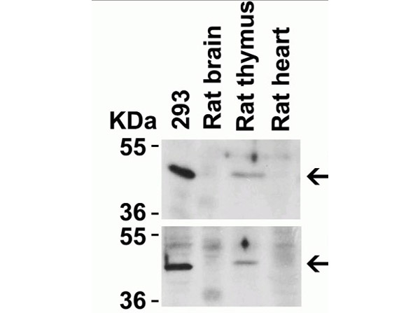
Animal Species Reactivity Western Blot Validation of CXCR4. Loading: 20 µg per lane. Lane 1: 293 lysate, Lane 2: Rat Brain lysate, Lane 3: Rat Thymus lysate, Lane 4: Rat Heart lysate. Primary Antibody: top panel Anti-CXCR4 (p/n 200-401-G91) at 2µg/mL, bottom panel (p/n 600-401-G92) at 2µg/mL for 1h at RT in 5% NFDM/TBST. Secondary: Goat anti-rabbit IgG HRP conjugate at 1:10000 dilution.
Immunofluorescence of CXCR4. Tissue: Rat Thymus. Fixation: 4% paraformaldehyde-fixed. Primary Antibody: CXCR4 at 20 µg/mL. Secondary: goat anti-rabbit IgG secondary antibody at 1/500 dilution (red) and DAPI staining (blue).
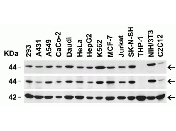
Western Blot of CXCR4. Load: 15 µg of lysates per lane. Primary Antibodies: TOP: human CXCR1 (n-term) (1 µg/mL), MIDDLE: human CXCR1 (internal) (p/n 600-401-G92) (1 µg/mL), and BOTTOM: beta-actin (1 µg/mL), for 1 h incubation at RT in 5% NFDM/TBST. Secondary: Goat anti-rabbit IgG HRP conjugate at 1:10000 dilution.
* Mehrwertsteuer und Versandkosten nicht enthalten. Irrtümer und Preisänderungen vorbehalten