
JAM A phospho Y280 Antibody, Rabbit, Polyclonal
Artikelnummer:
ROC-600-401-GN5S
- Bilder (8)

| Artikelname: | JAM A phospho Y280 Antibody, Rabbit, Polyclonal |
| Artikelnummer: | ROC-600-401-GN5S |
| Hersteller Artikelnummer: | 600-401-GN5S |
| Alternativnummer: | ROC-600-401-GN5S |
| Hersteller: | Rockland Immunochemicals |
| Wirt: | Rabbit |
| Kategorie: | Antikörper |
| Applikation: | ELISA, IF, WB |
| Spezies Reaktivität: | Human |
| Immunogen: | Affinity purified Anti-JAM A pY280 antibody was prepared from whole rabbit serum produced by repeated immunizations with a synthetic peptide corresponding to the c-term and phosphorylated at the tyrosine 280 position of Human JAM A protein. |
| Konjugation: | Unconjugated |
| Alternative Synonym: | rabbit anti-JAM A pY280 antibody, JAM-A, Junctional adhesion molecule A, JAM-1, Junctional adhesion molecule 1, Platelet F11 receptor, Platelet adhesion molecule 1, PAM-1, CD321, JAM1, JCAM, JAM 1, JAMA |
| Klonalität: | Polyclonal |
| Konzentration: | 1.0 mg/ml by UV absorbance at 280 nm |
| NCBI: | 058642 |
| UniProt: | 50848 |
| Puffer: | 0.02 M Potassium Phosphate, 0.15 M Sodium Chloride, pH 7.2 |
| Formulierung: | Liquid (sterile filtered) |
| Target-Kategorie: | Human |
| Antibody Type: | Primary Antibody |
| Application Verdünnung: | ELISA: 5 ug/ml, IF Microscopy: User optimized, WB: 1 ug/ml |
| Anwendungsbeschreibung: | This affinity purified antibody has been tested for use in ELISA, IF, and western blot. Specific conditions for reactivity should be optimized by the end user. Expect a band ~ 32.5 kDa in size corresponding to JAM A by western blotting in the appropriate |
|
|
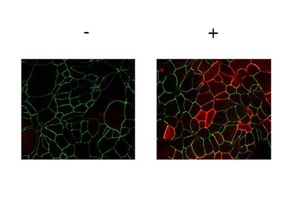
Immunofluorescence Microscopy of Rabbit anti-JAMA pY280 antibody. Tissue: T84 cells (untreated/treated). Fixation: 0.5% PFA. Antigen retrieval: not required. Primary antibody: JAMA pY280 antibody at 2 µg/mL for 1 hr at RT. Secondary antibody: Fluorescein rabbit secondary antibody at 1:10,000 for 45 min at RT. Localization: JAMA pY280 is along the cell membrane and cell junction. Staining: JAMA pY280 as red fluorescent signal. JCYIA |
|
|
|
|
|
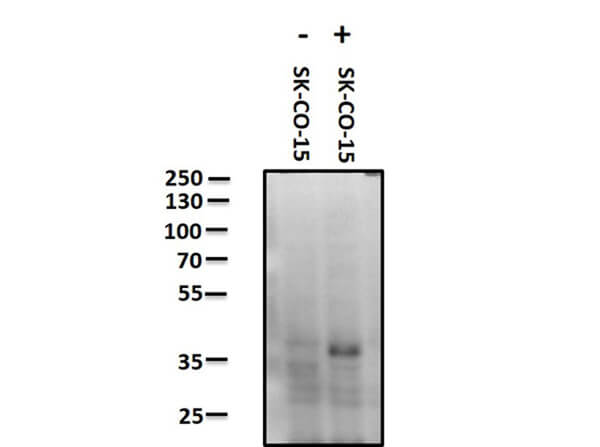
Western Blot of Rabbit anti-JAM A pY280 antibody. Lane 1: SK-CO-15 negative control. Lane 2: SK-CO-15 pervanadate treated positive control. Load: 10 µg per lane. Primary antibody: JAM A pY280 antibody at 1 ug/mL for overnight at 4C. Secondary antibody: Peroxidase rabbit secondary antibody at 1:40,000 for 30 min at RT. Block: MB-070 for 30 minutes at RT. Predicted/Observed size: ~ 32.5 kDa. JCYIA |
|
|
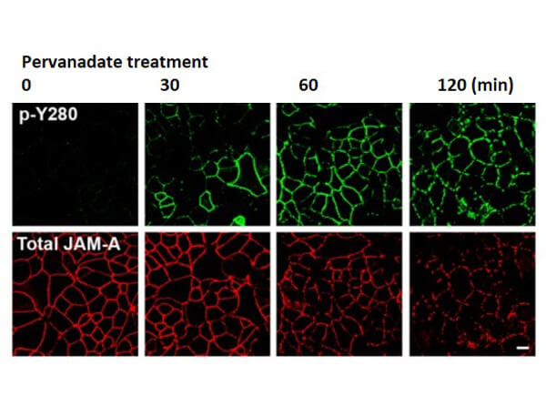
Confocal Immunofluorescence Microscopy of Rabbit Anti-JAM-A pY280 antibody of confluent (intestinal epithelial cells) IECs. Tissue: SK CO-15 cells. Treatment: pervanadate at time points 0, 30, 60, 120 mins. Fixation: 4% PFA. Permeabilization: 1% SDS. Costained Green: Anti-Phospho JAM-A Y280 Antibody, FITC conjugated secondary, Red: Anti-Total JAM-A, Alexa-conjugated secondary antibodies. Results: pervanadate treatment led to a time-dependent increase in phosphorylation of JAM-A Y280 that correlated with decreased localization of JAM-A at cell-cell contacts. Scale bar: 10 µm. See additional information in Mol Biol Cell. 2019 Mar 1, 30(5): 566-578. PMID: 30625033. |
|
|
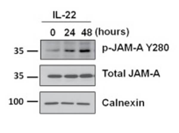
Western Blot of Rabbit Anti-JAM-A pY280 antibody with IL-22. Lysates: T84 cells. Treatment: hu rec. IL-22 at time points 0, 24, 48 hrs. Primary antibodies: JAM-A pY280, total JAM-A, or Calnexin. Calnexin was used as a loading control. Secondary antibody: horseradish peroxidase secondary antibody. Results: Exposure of IECs to other cytokines (IL-17A, IL-22, TNFalpha, or IFNgamma) results in tyrosine phosphorylation of JAM-A at Y280 and a leaky barrier. See additional information in Mol Biol Cell. 2019 Mar 1, 30(5): 566-578. PMID: 30625033. |
|
|
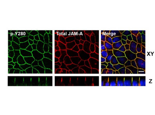
Confocal Immunofluorescence Microscopy of Rabbit Anti-JAM-A pY280 antibody in polarized epithelial cells. Tissue: T84 cells were grown on Transwell filters until confluent. Treatment: pervanadate. Fixation: 4% PFA. Permeabilization: 1% SDS. Costained Green: Anti-Phospho JAM-A Y280 Antibody, FITC conjugated secondary, Red: Anti-Total JAM-A, Alexa-conjugated secondary antibodies. Localization: tight junctions, seen in Confocal Z-stacks. Scale bar: 10 µm. See additional information in Mol Biol Cell. 2019 Mar 1, 30(5): 566-578. PMID: 30625033. |
|
|
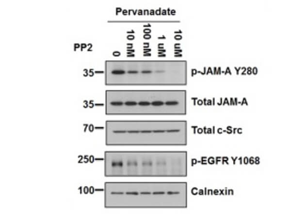
Western Blot of Rabbit Anti-JAM-A pY280 antibody with PP2. Lysates: T84 cells. Treatments: Pervanadate, PP2 at 0, 10nM, 100nM, 1µM, 10µM. Primary antibodies: p-JAM-A Y280, total JAM-A, total c-Src, p-EGFR Y1068, or Calnexin. p-EGFR Y1068 was used as a positive control for PP2. Calnexin was used as a loading control. Secondary antibody: horseradish peroxidase secondary antibody. Results: PP2 dose-dependent decrease in tyrosine phosphorylation of JAM-A Y280 following pervanadate treatment. See additional information in Mol Biol Cell. 2019 Mar 1, 30(5): 566-578. PMID: 30625033. |
|
|
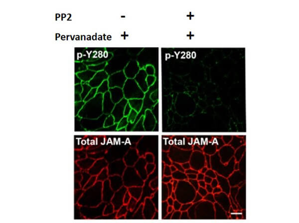
Immunofluorescence Microscopy of Rabbit Anti-JAM-A pY280 antibody. Tissue: T84 cells. Pretreatment: PP2. Treatment: Pervanadate. Fixation: 4% PFA. Permeabilization: 1% SDS. Costained Green: Anti-Phospho JAM-A Y280 Antibody, FITC conjugated secondary, Red: Anti-Total JAM-A, Alexa-conjugated secondary antibodies. Results: The Src family kinase inhibitor PP2 inhibits pervanadate-dependent phosphorylation of JAM-A Y280, as they reported to modulate tyrosine phosphorylation of junctional proteins and influence epithelial barrier function. See additional information in Mol Biol Cell. 2019 Mar 1, 30(5): 566-578. PMID: 30625033. |
Produktgarantie und fachkundiger Support
