Anti-ZO-1 antibody was prepared from whole rabbit serum produced by repeated immunizations with a synthetic peptide corresponding to an internal portion of human ZO-1 conjugated to Keyhole Limpet Hemocyanin (KLH).
Konjugation:
Unconjugated
Alternative Synonym:
rabbit anti-ZO-1 antibody, ZO 1, ZO1, Tight junction protein ZO-1, Tight junction protein 1, Zona occludens protein 1, Zonula occludens protein 1, TJP1
0.02 M Potassium Phosphate, 0.15 M Sodium Chloride, pH 7.2
Formulierung:
Liquid (sterile filtered)
Target-Kategorie:
Human
Antibody Type:
Primary Antibody
Application Verdünnung:
ELISA: 10,000-1:50,000, Flow Cytometry: User Optimized, IHC: 1:100 - 1:200, IF Microscopy: 10 µg/ml, WB: 1:1000
Anwendungsbeschreibung:
Anti-ZO-1 Antibody has been tested in Western Blot, ELISA, Immunohistochemistry, Immunofluorescence, and Flow Cytometry. Expect a band at ~245 and/or 195.5 kDa in western blot using appropriate lysates. Positive control whole cell lysates used A549 and P
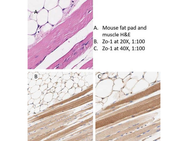
Immunohistochemistry of Rabbit Anti-ZO-1 antibody. Tissue: mouse adipose tissue and muscle. Fixation: formalin fixed paraffin embedded. Antigen retrieval: heat induced (HIER) using Citrate Buffer for 20min. Primary antibody: ZO-1 antibody at 1:100 for 30min at RT. Secondary Antibody: Anti-Rabbit Poly-HRP-IgG Ready-to-Use for 8min at RT. Localization: ZO-1 will stain cell-cell junctions. Staining: DAB. Counter Stain: Hematoxylin.
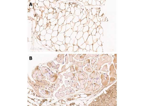
Immunohistochemistry of Rabbit anti-ZO-1 antibody. Tissue: mouse adipose tissue. Fixation: formalin fixed paraffin embedded. Epitope retrieval: heat induced (HIER). Primary antibody: ZO-1 antibody at 1:100 [A] and 1:200 [B] for 1 h at RT. Localization: ZO-1 will stain cell-cell junctions. Visualized with WARP RED on MACH 4 universal AP polymer detection system.
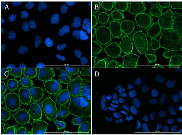
Immunofluorescence Microscopy of Rabbit anti-ZO-1 antibody. Tissue: CaCO2. Fixation: 4% PFA. Permeabilization: 0.3%Triton X-100. Primary antibody: ZO-1 antibody at 15µg/mL overnight at 2-8C. Secondary antibody: Donkey Anti-Rabbit IgG DyLight(TM) 488 Conjugated Preadsorbed (p/n 611-741-127) at 5µg/mL for 1 h at RT. Localization: membrane. Staining: (A)DAPI. (B)ZO-1+DyLight488. (C)Merge A-B. (D) Secondary Only.
Immunofluorescence Microscopy of Rabbit anti-ZO-1 antibody. Tissue: Caco2. Fixation: 0.5% PFA [A,C]. 0.5% MeOH [B,D]. Antigen retrieval: not required. Primary antibody: ZO-1 antibody at 10 µg/mL for 1 h at RT. Secondary antibody: Anti-RABBIT IgG DyLight(TM) 488 Conjugated Preadsorbed (p/n 611-741-127) at 5 ug/ml for 1 h at RT. Localization: (1) most epithelial cell junctions, (2) both in endothelial cells and the highly specialized epithelial junctions of renal and Sertoli cells. Staining: Target as green fluorescent signal with DAPI (blue) nuclear counterstain.
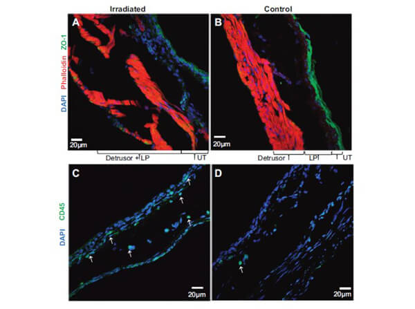
Immunofluorescence of ZO-1 and CD45. Representative confocal images of irradiated group (A) and controls (B) demonstrate a dramatic reduction of green fluorescence for ZO-1 in urothelium layer of irradiated group. Bladder sections were labeled with anti-ZO-1 antibodies and secondary donkey antibody Alexa 488 to localize the tight junction protein ZO-1 and with rhodamine-phalloidin to label the actin cytoskeleton (red) and DAPI to label the nuclei. Decreased expression of ZO-1 in urothelium layer of irradiated group was associated with an increase in CD45-positive, green fluorescent leukocytes (indicated by whit
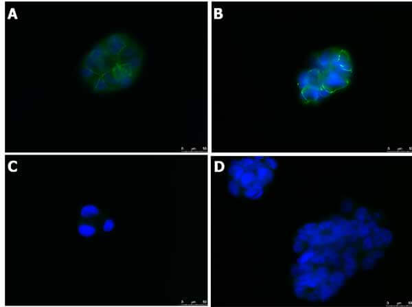
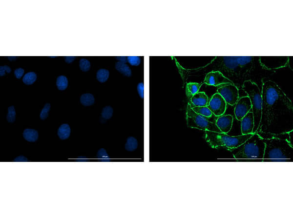
Immunofluorescence microscopy of Anti-ZO-1 in Caco-2 cells using FITC-conjugated Fluorescent TrueBlot anti-rabbit IgG (p/n 18-0216-32) for detection. Caco-2 cells were fixed with 4% PFA, blocked (5% mouse serum/0.3% Triton X-100 in 1X PBS) for 1hr, then incubated with 15µg/mL of anti-ZO-1 primary antibody (Cat. No. 600-401-GU7) at 4C overnight. Following 3 washes in 1X PBS for 5min each, 5µg/mL of FITC-conjugated Fluorescent TrueBlot anti-rabbit IgG was added and allowed to incubate for 1hr at room temperature. Nuclei were counterstained with DAPI present in mounting medium. Predicted cell localization is cell membrane and cell junctions. Image taken at 40X magnification. (Right) Merged DAPI (blue)/ZO-1 (green), image shown (Left) secondary antibody only.
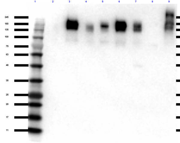
Western Blot of Rabbit Anti-ZO-1 Antibody. Lane 1: Opal Pre-stained MW ladder (p/n MB-210-0500). Lane 2: WM-115 Whole Cell Lysate (p/n W09-001-GY8). Lane 3: A549 Whole Cell Lysate (p/n W09-001-372). Lane 4: HeLa Whole Cell Lysate (p/n W09-000-364). Lane 5: HeLa Whole Cell Lysate CCCP Stimulated (p/n W09-001-GZ0). Lane 6: PC-3 Whole Cell Lysate (p/n W09-001-GV6). Lane 7: SK-OV-3 Whole Cell Lysate (p/n W09-001-GX9). Lane 8: Mouse Testis Lysate (p/n W10-000-GZ2). Lane 9: Rat Testis Lysate (p/n W12-000-GZ3). Load: 10 µg per lane. Primary antibody: ZO-1 antibody at 1:1000 for overnight at 4C. Secondary antibody: rabbit secondary HRP antibody (p/n 611-103-122) at 1:70,000 for 1 hr at RT. Block: 5% BLOTTO (p/n B501-0500) for 30 min at RT. Predicted/Observed size: ~187, 195 kDa for ZO-1.
* Mehrwertsteuer und Versandkosten nicht enthalten. Irrtümer und Preisänderungen vorbehalten