RIP3 Antibody was produced from whole rabbit serum prepared by repeated immunizations with a peptide corresponding to 14 amino acids near the carboxy terminus of murine RIP3. The immunogen is located within the last 50 amino acids of RIP3.
Konjugation:
Unconjugated
Alternative Synonym:
Receptor-interacting serine/threonine-protein kinase 3, RIP-like protein kinase 3, Receptor-interacting protein 3, RIP-3, mRIP3, Ripk3, Rip3
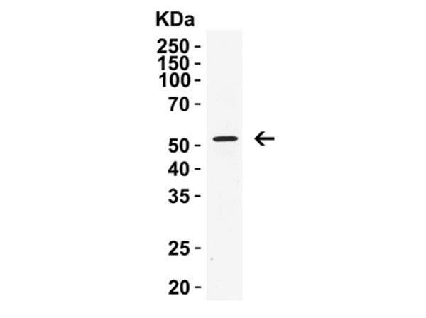
Anti-Rip3 Antibody is tested for use in E, WB, IF, IP, and IHC. Expect a band approximately ~53 kDa for mouse and ~57kDa for human RIP3, on specific lysates or tissues. Observed: 53kD for mouse RIP3 and 57kD for human RIP3. Specific conditions for reacti
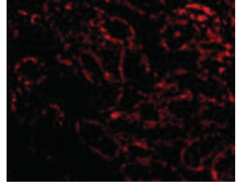
Immunofluorescence Microscopy of RIP3 antibody. Tissue: Rat kidney cells. Fixation: 0.5% PFA. Antigen retrieval: not required. Primary antibody: RIP3 antibody at 20 µg/mL for 1 h at RT. Secondary antibody: Fluorescein rabbit secondary antibody at 1:10,000 for 45 min at RT. Localization: RIP3 is cytoplasmic and is also localized in the cell membrane and mitochondria. Staining: RIP3 as red fluorescent signal.
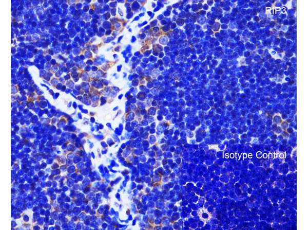
Immunohistochemistry Validation of RIP3. Tissue: Rat Thymus. Fixation: paraffin-embedded, formaldehyde and blocked with 10% serum for 1 h at RT. Antigen retrieval: heat mediation with a citrate buffer (pH6). Primary Antibody: anti-RIP3 antibody at 1 µg/ml overnight at 4C. Secondary: goat anti-rabbit IgG H&L (HRP) at 1:250. Counter stained with Hematoxylin.
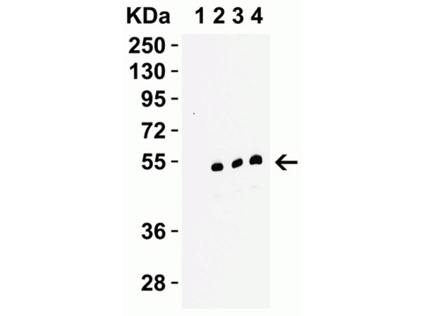
Western Blot Validation of RIP3. Load: 15 µg of mouse C2C12 Cell lysates per lane. Primary antibody: RIP3 at (Lane 1: 0.1 µg/mL in the presence of peptide blocking. Lane 2: 0.1 µg/mL. Lane 3: 0.2 µg/mL. Lane 4: 0.5 µg/mL) for 1h incubation at RT in 5% NFDM/TBST. Secondary: Goat anti-rabbit IgG HRP conjugate at 1:10000 dilution.
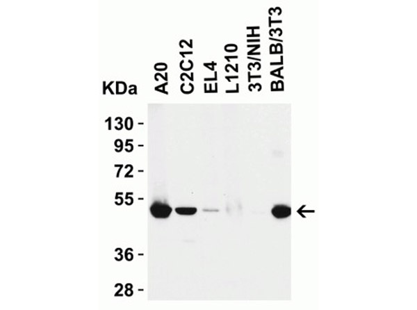
Western Blot Validation of RIP3. Load: 15 µg of mouse cell lysates. Lane 1: A20, Lane 2: C2C12, Lane 3: EL4, Lane 4: L1210, Lane 5: 3T3/NIH, Lane 6: BALB/3T3. Primary antibody: RIP3 at 0.5 µg/mL for 1h incubation at RT in 5% NFDM/TBST. Secondary: Goat anti-rabbit IgG HRP conjugate at 1:10000 dilution.
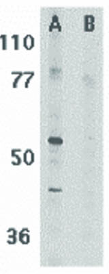
Western Blot of RIP3 antibody. Lane 1: Mouse 3T3 whole cell lysate. Lane 2: Mouse 3T3 whole cell lysate in the presence of blocking peptide. Load: 35 µg per lane. Primary antibody: RIP3 antibody at 1 µg/mL for overnight at 4C. Secondary antibody: Peroxidase rabbit secondary antibody at 1:10,000 for 45 min at RT. Block: 5% BLOTTO overnight at 4C. Predicted/Observed size: 56.9 kDa, 62 kDa for RIP3. Other band(s):RIP3 splice variants and isoforms.
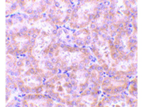
Immunohistochemistry of RIP3 antibody. Tissue: Rat kidney tissue. Fixation: formalin fixed paraffin embedded. Antigen retrieval: not required. Primary antibody: RIP3 antibody at 5 µg/mL for 1 h at RT. Secondary antibody: Peroxidase rabbit secondary antibody at 1:10,000 for 45 min at RT. Localization: RIP3 is cytoplasmic and is also localized in the cell membrane and mitochondria. Staining: RIP3 as precipitated brown signal with hematoxylin purple nuclear counterstain.
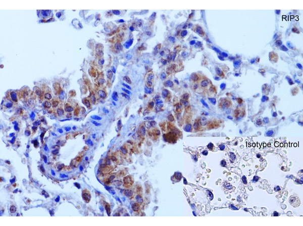
Immunohistochemistry Validation of RIP3 in Mouse Lung.
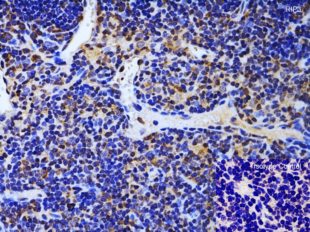
Immunohistochemistry Validation of RIP3. Tissue: Mouse Thymus. Fixation: paraffin-embedded, formaldehyde and blocked with 10% serum for 1 h at RT. Antigen retrieval: heat mediation with a citrate buffer (pH6). Primary Antibody: anti-RIP3 antibody at 1 µg/ml overnight at 4C. Secondary: goat anti-rabbit IgG H&L (HRP) at 1:250. Counter stained with Hematoxylin.
Western Blot Validation of RIP3. Load: 15 µg of Rat Thymus lysate. Primary antibody: RIP3 at 0.5 µg/mL for 1h incubation at RT in 5% NFDM/TBST. Secondary: Goat anti-rabbit IgG HRP conjugate at 1:10000 dilution.
* Mehrwertsteuer und Versandkosten nicht enthalten. Irrtümer und Preisänderungen vorbehalten