ST2 Antibody was produced from whole rabbit serum prepared by repeated immunizations with a synthetic peptide corresponding to amino acids at the n-terminus of mouse ST2. This peptide is common to all three known ST2 isoforms.
Konjugation:
Unconjugated
Alternative Synonym:
Ly84, St2, Ste2, Interleukin-1 receptor-like 1, Interleukin-33 receptor alpha chain1, Lymphocyte antigen 84, Protein ST2, Protein T1
0.02 M Potassium Phosphate, 0.15 M Sodium Chloride, pH 7.2
Formulierung:
Liquid (sterile filtered)
Target-Kategorie:
Mouse
Antibody Type:
Primary Antibody
Application Verdünnung:
WB: 1-2 ug/mL
Anwendungsbeschreibung:
Anti-ST2 Antibody has been tested in ELISA, WB, IF, and IHC. Expect a band approximately ~64.8kDa on specific lysates. Specific conditions for reactivity should be optimized by the end user.
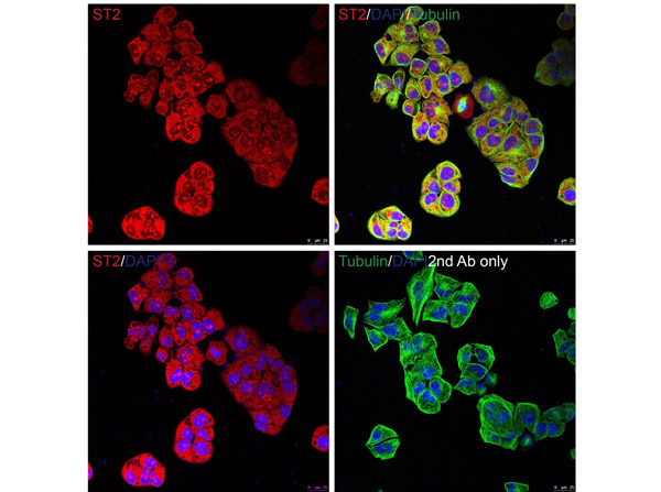
Immunofluorescence of ST2. Cell: HeLa Cells.Fixation: PFA-fixed, Primary Antibody: Anti-ST2 at 20 µg/mL.Secondary: goat anti-rabbit IgG secondary antibody at 1:1000 dilution (red) and DAPI staining (blue). Alpha tubulin was stained with anti-alpha tubulin antibody following by goat anti-mouse IgG secondary antibody (green). Images were captured with confocal microscopy.
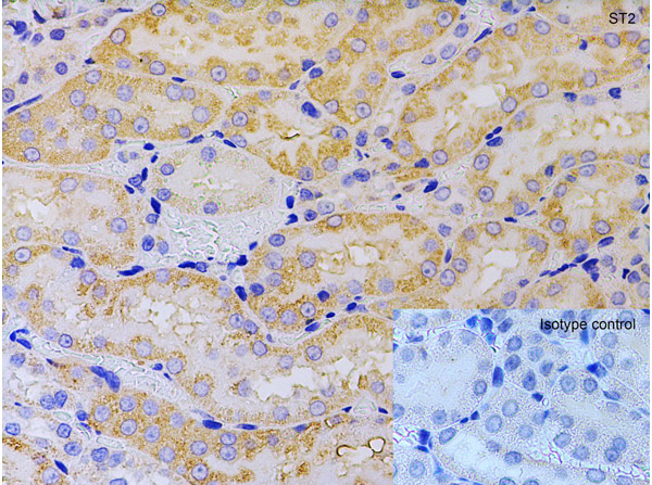
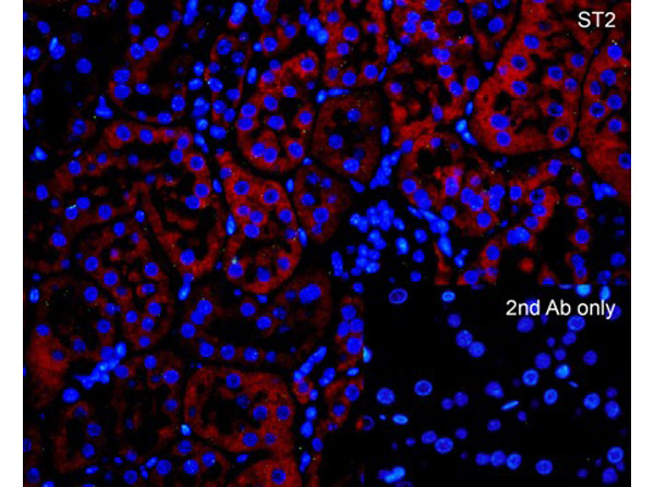
Immunohistochemistry of ST2. Tissue: rat kidney tissue. Fixation: paraffin-embedded, formaldehyde and blocked with 10% serum for 1 h at RT. Antigen retrieval: heat mediation with a citrate buffer (pH6). Priamry Antibody: anti-ST2 antibody at 5 µg/ml overnight at 4C. Secondary: goat anti-rabbit IgG H&L (HRP) at 1:250. Counter stained with Hematoxylin.
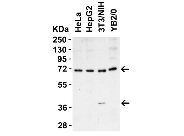
Western Blot of ST2.Load: 15 µg of Human, Mouse, or Rat lysate per lane. Lane 1: HeLa, Lane 2: HepG2, LAne 3: 3T3/NIH, Lane 4: YB2/0. Primary Antibody: anti-ST2 at 1 µg/mL for 1h at RT in 5% NFDM/TBST. Secondary: Goat Anti-Rabbit IgG HRP conjugate at 1:10000 dilution.
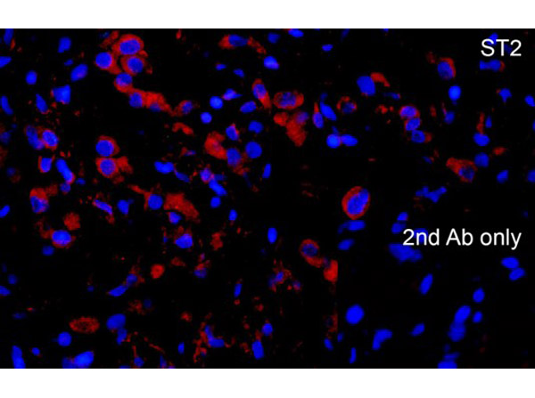
Immunofluorescence of ST2. Tissue: Human Lung tissue. Fixation: PFA-fixed. Primary Antibody: Anti-ST2 at 10 µg/mL. Secondary: goat anti-rabbit IgG secondary antibody at 1:1000 dilution (red) and DAPI staining (blue).
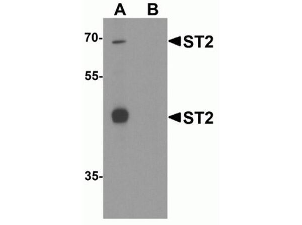
Western Blot of ST2. Load: 293 cell lysate.Primary Antibody: Anti-ST2 at 1µg/mL for 1h at RT in 5% NFDM/TBST. Lane A: In the absence of blocking peptide Lane B: In the presence of blocking peptide. Secondary: Goat anti-rabbit IgG HRP conjugate at 1:10000 dilution.
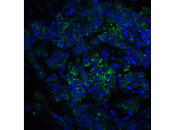
Immunofluorescence of ST2. Tissue: Human Lung Cancer Tissue. Fixation: 4% paraformaldehyde-fixed. Primary Antibody: Anti-ST2 at 5 µg/mL. Secondary: goat anti-rabbit IgG secondary antibody at 1:500 dilution (green) DAPI staining (blue).
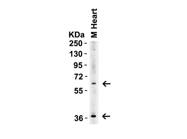
Western Blot of ST2. Load: 15 µg of Mouse Heart lysate. Primary antibody: anti-ST2 at 1µg/mL for 1h at RT in 5% NFDM/TBST. Secondary: Goat anti-rabbit IgG HRP conjugate at 1:10000 dilution.
Immunofluorescence of ST2. Tissue: mouse kidney tissueFixation: PFA-fixed. Primary Antibody: Anti-ST2 at 10 µg/mL. Secondary: goat anti-rabbit IgG secondary antibody at 1:1000 dilution (red) and DAPI staining (blue).
* Mehrwertsteuer und Versandkosten nicht enthalten. Irrtümer und Preisänderungen vorbehalten