TSLP Antibody was produced from whole rabbit serum prepared by repeated immunizations with a peptide corresponding to an internal region of human TSLP.
0.02 M Potassium Phosphate, 0.15 M Sodium Chloride, pH 7.2
Formulierung:
Liquid (sterile filtered)
Target-Kategorie:
Human
Antibody Type:
Primary Antibody
Application Verdünnung:
IHC: 1-2.5µg/mL, IF Microscopy: 20µg/mL, WB: 0.25-4µg/mL
Anwendungsbeschreibung:
Anti-TSLP Antibody is tested for use in E, WB, IF, and IHC. Expect a band approximately ~18.1 kDa on specific lysates. Western Blot, Immunohistochemistry, and Immunofluorescence tested in human and mouse samples, Immunocytochemistry in human samples. Spe
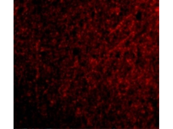
Immunofluorescence of TSLP. Tissue: Human Brain Tissue. Fixation: 4% paraformaldehyde-fixed. Primary Antibody: Anti-TSLP at 20 µg/mL. Secondary Antibody: goat anti-rabbit IgG secondary antibody at 1:500 dilution (red).
Immunohistochemistry of TSLP. Tissue: Human Brain Tissue. Fixation: paraffin-embedded, formaldehyde and blocked with 10% serum for 1 h at RT. Antigen retrieval was by heat mediation with a citrate buffer (pH6). Primary Antibody: anti-TSLP antibody at 2.5 µg/ml overnight at 4C. Secondary: goat anti-rabbit IgG H&L (HRP) at 1:250. Counter stained with Hematoxylin.
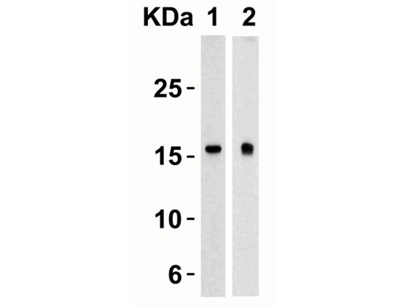
Western Blot of TSLP. Load: 15 µg of lysates per lane Human Heart (Lane 1) and Human Prostate (Lane 2). Primary antibody: Anti-TSLP at 4 µg/mL for 1h incubation at RT in 5% NFDM/TBST. Secondary: Goat anti-rabbit IgG HRP conjugate at 1:10000 dilution.
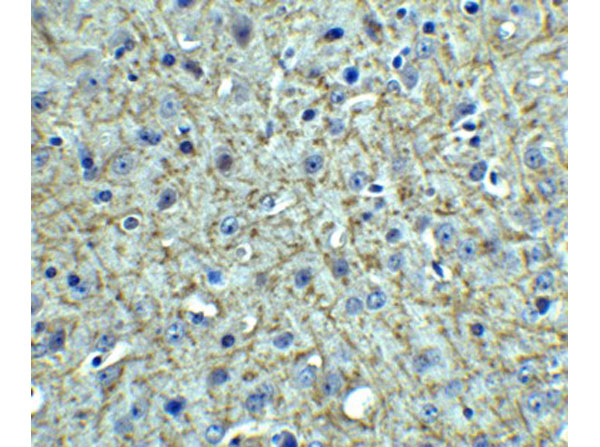
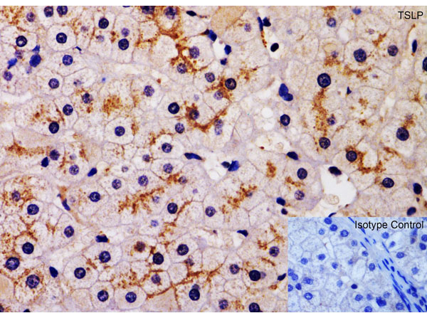
Immunohistochemistry of TSLP. Tissue: Human Liver Tissue. Fixation: paraffin-embedded, fixed with formaldehyde and blocked with 10% serum for 1 h at RT. Antigen retrieval: heat mediation with a citrate buffer (pH6). Primary Antibody: anti-TSLP antibody at 2 µg/mL overnight at 4CSecondary: goat anti-rabbit IgG H&L (HRP) at 1:250. Counter stained with Hematoxylin.
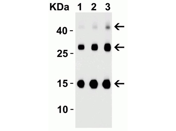
Western Blot of TSLP. Load: 30ng of TSLP partial human recombinant protein per lane. Primary antibody: Anti-TSLP at (Lane 1: 0.25 µg/mL, Lane 2: 0.5 µg/mL, Lane 3: 1 µg/mL) for 1h incubation at RT in 5% NFDM/TBST. Secondary: Goat anti-rabbit IgG HRP conjugate at 1:10000 dilution. Observed MW: TLSP partial human recombinant protein 15kD, the observed bands at 30kD and 45kD are the dimer and trimer, respectively.
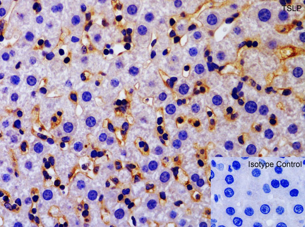
Immunohistochemistry of TSLP. Tissue: Mouse Liver Tissue. Fixation: paraffin-embedded, fixed with formaldehyde and blocked with 10% serum for 1 h at RT. Antigen retrieval: heat mediation with a citrate buffer (pH6). Primary Antibody: anti-TSLP antibody at 2 µg/mL overnight at 4CSecondary: goat anti-rabbit IgG H&L (HRP) at 1:250. Counter stained with Hematoxylin.
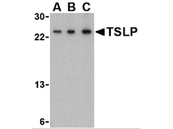
Western Blot of TSLP. Load: 15 µg of Mouse A-20 Cell Line lysates per lane. Primary antibody: Anti-TSLP at (A: 0.5 µg/mL, B: 1 µg/mL, C: 2 µg/mL) for 1h incubation at RT in 5% NFDM/TBST. Secondary: Goat anti-rabbit IgG HRP conjugate at 1:10000 dilution.
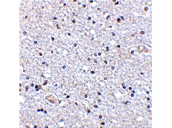
Immunohistochemistry of TSLP. Tissue: Human Brain Tissue. Fixation: paraffin-embedded, fixed with formaldehyde and blocked with 10% serum for 1 h at RT. Antigen retrieval: heat mediation with a citrate buffer (pH6). Primary Antibody: anti-TSLP antibody at 2.5 µg/mL overnight at 4CSecondary: goat anti-rabbit IgG H&L (HRP) at 1:250. Counter stained with Hematoxylin.
* Mehrwertsteuer und Versandkosten nicht enthalten. Irrtümer und Preisänderungen vorbehalten